The heart, an essential muscular pump, ensures the circulation of bloodBlood is composed of red blood cells, white blood cells, platelets, and plasma. Red blood cells are responsible for transporting oxygen and carbon dioxide. White blood cells make up our immune defense system. Platelets contribute to blood throughout the body, acting as the engine that sustains life.
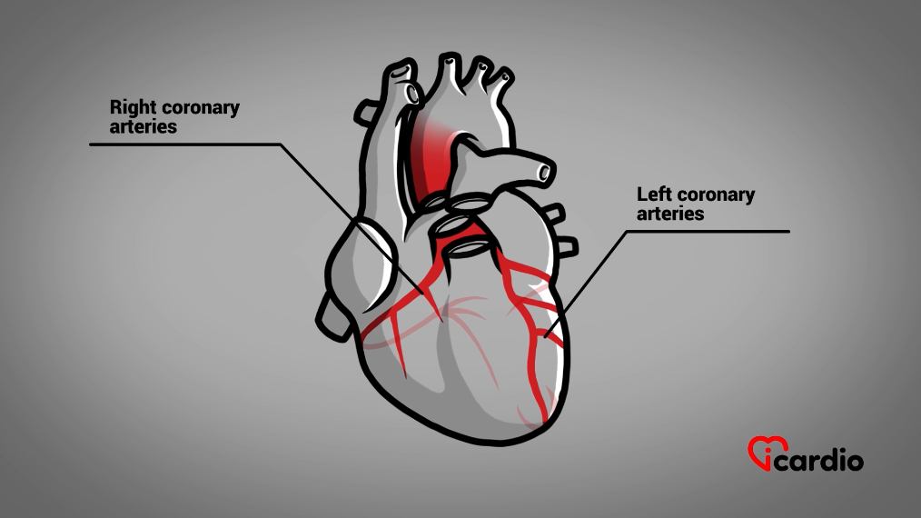
The coronary arteries
This vital muscle functions thanks to the supply of oxygen delivered by a network of bloodBlood is composed of red blood cells, white blood cells, platelets, and plasma. Red blood cells are responsible for transporting oxygen and carbon dioxide. White blood cells make up our immune defense system. Platelets contribute to blood vessels called coronary arteriesThe two coronary arteries, the right and the left, form the blood network that supplies the heart with oxygen and nutrients. They are located directly on the surface of the heart and branch into smaller vessels that. These arteries surround and penetrate the heart, delivering the oxygen it needs to function properly. Their diameter ranges from 2 to 4 mm.
Any reduction in the size of these arteries, even slight, can impair the oxygen supply to the heart muscle, compromising its function. If the reduction becomes significant enough, it can lead to irreversible damage to the heart.
There are three acute alterations that can obstruct bloodBlood is composed of red blood cells, white blood cells, platelets, and plasma. Red blood cells are responsible for transporting oxygen and carbon dioxide. White blood cells make up our immune defense system. Platelets contribute to blood flow in the coronary arteriesThe two coronary arteries, the right and the left, form the blood network that supplies the heart with oxygen and nutrients. They are located directly on the surface of the heart and branch into smaller vessels that, all originating from the muscular layer called the media:
1- Artery spasm: This phenomenon can occur spontaneously or be triggered by substances such as cocaine.
It involves a contraction of the media layer, narrowing the bloodBlood is composed of red blood cells, white blood cells, platelets, and plasma. Red blood cells are responsible for transporting oxygen and carbon dioxide. White blood cells make up our immune defense system. Platelets contribute to blood vessel. The severity of the spasm, whether partial or complete, along with its duration, determines its impact.
This temporary blockage can have serious consequences if the oxygen supply to the heart is obstructed for too long.
2- Spontaneous dissection: This is a tear in the media, the cause of which is still poorly understood. This rupture may be linked to a weakness or abnormality in the artery wall.
As with a spasm, the effects depend on the extent of the tear, ranging from a partial obstruction to a total blockage of the vessel.
Plus souvent désignée comme étant une rupture de plaque de cholestérol, c’est de loin la cause la plus fréquente d’altération de la circulation coronaire.
Let’s take a closer look at the last case.
Cholesterol Plaque
The rupture of an atheroma plaque is the most common cause of acute coronary arteryThe two coronary arteries, the right and the left, form the blood network that supplies the heart with oxygen and nutrients. They are located directly on the surface of the heart and branch into smaller vessels that obstruction.
These plaques, mainly composed of cholesterolCholesterol is essential for the proper functioning of the human body, but it can also have harmful effects if present in excess. >>, gradually accumulate in the media of the arteries.
At some point, the plaque may rupture, triggering the formation of a clot that disrupts bloodBlood is composed of red blood cells, white blood cells, platelets, and plasma. Red blood cells are responsible for transporting oxygen and carbon dioxide. White blood cells make up our immune defense system. Platelets contribute to blood flow.
When an atheroma plaque ruptures, the body responds by triggering a repair mechanism to seal the breach.
This process begins with the activation of bloodBlood is composed of red blood cells, white blood cells, platelets, and plasma. Red blood cells are responsible for transporting oxygen and carbon dioxide. White blood cells make up our immune defense system. Platelets contribute to bloodplateletsPlatelets, also known as thrombocytes, are small cell fragments produced by the bone marrow. They play a crucial role in blood clotting by forming aggregates and clots to stop bleeding when blood vessels are damaged. >>, which play a critical role in bloodBlood is composed of red blood cells, white blood cells, platelets, and plasma. Red blood cells are responsible for transporting oxygen and carbon dioxide. White blood cells make up our immune defense system. Platelets contribute to blood clotting.
It's "911" for Platelets
PlateletsPlatelets, also known as thrombocytes, are small cell fragments produced by the bone marrow. They play a crucial role in blood clotting by forming aggregates and clots to stop bleeding when blood vessels are damaged. >> are tiny particles circulating in the bloodBlood is composed of red blood cells, white blood cells, platelets, and plasma. Red blood cells are responsible for transporting oxygen and carbon dioxide. White blood cells make up our immune defense system. Platelets contribute to blood, originating from large cell fragments found in the bone marrow. Their primary function is to assist in bloodBlood is composed of red blood cells, white blood cells, platelets, and plasma. Red blood cells are responsible for transporting oxygen and carbon dioxide. White blood cells make up our immune defense system. Platelets contribute to blood coagulation, a process that stops bleeding by sealing breaks in bloodBlood is composed of red blood cells, white blood cells, platelets, and plasma. Red blood cells are responsible for transporting oxygen and carbon dioxide. White blood cells make up our immune defense system. Platelets contribute to blood vessels.
Understanding Blood Clotting "101"
When someone gets a cut, bloodBlood is composed of red blood cells, white blood cells, platelets, and plasma. Red blood cells are responsible for transporting oxygen and carbon dioxide. White blood cells make up our immune defense system. Platelets contribute to blood flows out due to a breach in the bloodBlood is composed of red blood cells, white blood cells, platelets, and plasma. Red blood cells are responsible for transporting oxygen and carbon dioxide. White blood cells make up our immune defense system. Platelets contribute to blood vessels. To stop the bleeding, the body sends out a distress signal, like an emergency call. BloodBlood is composed of red blood cells, white blood cells, platelets, and plasma. Red blood cells are responsible for transporting oxygen and carbon dioxide. White blood cells make up our immune defense system. Platelets contribute to blood clotting is the natural process that steps in to seal the wound.
Formation of the Blood Clot
Platelets are the first responders to this emergency call. When they come into contact with the inner layers of the vessel, called the intima, they change shape, develop tentacle-like structures, and begin clumping together. These clumping forms the foundation of the clot.
Simultaneously, the plateletsPlatelets, also known as thrombocytes, are small cell fragments produced by the bone marrow. They play a crucial role in blood clotting by forming aggregates and clots to stop bleeding when blood vessels are damaged. >> release substances that attract more plateletsPlatelets, also known as thrombocytes, are small cell fragments produced by the bone marrow. They play a crucial role in blood clotting by forming aggregates and clots to stop bleeding when blood vessels are damaged. >> and trigger the transformation of certain bloodBlood is composed of red blood cells, white blood cells, platelets, and plasma. Red blood cells are responsible for transporting oxygen and carbon dioxide. White blood cells make up our immune defense system. Platelets contribute to bloodproteinsProteins are fundamental components manufactured by the cells of our body. They play an essential role in many biological functions, acting as hormones, antibodies, and even cholesterol transporters, among others. >> into filaments.
These filaments form a mesh around the plateletsPlatelets, also known as thrombocytes, are small cell fragments produced by the bone marrow. They play a crucial role in blood clotting by forming aggregates and clots to stop bleeding when blood vessels are damaged. >> and red bloodBlood is composed of red blood cells, white blood cells, platelets, and plasma. Red blood cells are responsible for transporting oxygen and carbon dioxide. White blood cells make up our immune defense system. Platelets contribute to blood cells, reinforcing the structure of the clot and making it more secure to seal the breach effectively.
It's the Same Process in the Coronary Artery
When a cholesterolCholesterol is essential for the proper functioning of the human body, but it can also have harmful effects if present in excess. >> plaque ruptures in a coronary arteryThe two coronary arteries, the right and the left, form the blood network that supplies the heart with oxygen and nutrients. They are located directly on the surface of the heart and branch into smaller vessels that, the same coagulation process is triggered to seal the breach. PlateletsPlatelets, also known as thrombocytes, are small cell fragments produced by the bone marrow. They play a crucial role in blood clotting by forming aggregates and clots to stop bleeding when blood vessels are damaged. >> gather and form a clot to repair the damage, just as they would in the case of a cut.
However, in the coronary arteriesThe two coronary arteries, the right and the left, form the blood network that supplies the heart with oxygen and nutrients. They are located directly on the surface of the heart and branch into smaller vessels that, the formation of this clot can have serious consequences. It can result in either a partial or complete obstruction of the vessel.
In both cases, this disrupts the oxygen supply to the heart muscle, potentially leading to major complications, such as unstable angina or, in more severe cases, a myocardial infarction (heart attack).
Complete Coronary Artery Obstruction - STEMI
When a blood clot completely blocks a coronary artery due to the rupture of an atherosclerotic plaque, the heart muscle is deprived of oxygen and begins to suffer intensely. This condition is commonly referred to as a “heart attack,” medically known as STEMI (ST-Elevation Myocardial Infarction).
The formation of a clot in the coronary arteriesThe two coronary arteries, the right and the left, form the blood network that supplies the heart with oxygen and nutrients. They are located directly on the surface of the heart and branch into smaller vessels that is a dynamic process. Just as our body has a coagulation system to seal breaches in bloodBlood is composed of red blood cells, white blood cells, platelets, and plasma. Red blood cells are responsible for transporting oxygen and carbon dioxide. White blood cells make up our immune defense system. Platelets contribute to blood vessels, it also has a mechanism for dissolving clots.
A medical emergency where every minute counts
From the moment symptoms appear, a race against time begins. Without oxygen, heart muscle cells start to die quickly. The longer the obstruction remains, the greater and more irreversible the damage.
A part of the heart stops functioning
Unlike some other organs, the heart cannot regenerate dead cells after a myocardial infarction. The affected area loses its ability to contract, weakening the heart’s capacity to pump bloodBlood is composed of red blood cells, white blood cells, platelets, and plasma. Red blood cells are responsible for transporting oxygen and carbon dioxide. White blood cells make up our immune defense system. Platelets contribute to blood effectively.
The extent of the damage depends on the size of the oxygen-deprived area and how quickly medical intervention is provided.
How to Recognize a Heart Attack?
Heart attack symptoms can vary from person to person, but they generally have the following characteristics:
Intense and persistent chest pain: A crushing discomfort or tightness in the chest lasting more than 30 minutes.
Associated symptoms: Nausea, vomiting, shortness of breath, cold sweats, and a feeling of weakness.
Pain radiating elsewhere: Discomfort may extend to the left arm, sometimes to the right arm, jaw, or back.
The false impression of severe indigestion
In some cases, heart attack symptoms can mimic those of severe indigestion, delaying the patient’s reaction. When in doubt, it is always best to call 911 immediately.
Small Warning Signs: Do Not Ignore Them!
In the days leading up to a heart attack, some people experience mild chest discomfort that may seem insignificant.
Pain during activity or at rest: These discomforts are brief but recurrent and can occur even without physical exertion.
A sign of an underlying issue: These symptoms often indicate an ongoing clot formation in the coronary arteryThe two coronary arteries, the right and the left, form the blood network that supplies the heart with oxygen and nutrients. They are located directly on the surface of the heart and branch into smaller vessels that.
These warning pains are often a sign of unstable angina, a condition that can quickly develop into a heart attack. It is essential to consult a healthcare professional as soon as possible to prevent a major cardiac event.
The Heart’s Electrical Risk
A heart attack does not just damage the heart muscle; it can also disrupt the heart’s electrical system, leading to dangerous palpitations“Palpitation” is a symptom related to an abnormality in heartbeats. There are several types of arrhythmias. This term is like a surname that encompasses several first names. that may cause cardiac arrest.
Too Many Deaths Before Reaching the Hospital
Many heart attack victims do not survive long enough to receive medical care. Coronary arteryThe two coronary arteries, the right and the left, form the blood network that supplies the heart with oxygen and nutrients. They are located directly on the surface of the heart and branch into smaller vessels that disease remains one of the leading causes of sudden death.
It is common for patients to delay seeking medical attention, hoping the pain will subside on its own. Every minute counts—the sooner a diagnosis is made, the greater the chances of minimizing damage and saving a life.
Emergency responders act immediately
In major urban centers, paramedics perform an electrocardiogram (ECG) upon arrival to detect a heart attack. This rapid initial assessment helps direct the patient to the appropriate treatment without delay.
Direct transport to a specialized center
If the ECG confirms a heart attack, the patient is immediately taken to a specialized hospital where a coronary angioplasty can be performed to unblock the obstructed artery.
Initial treatment in the ambulance
Paramedics administer aspirin as soon as they take charge of the patient. This medication prevents blood plateletsPlatelets, also known as thrombocytes, are small cell fragments produced by the bone marrow. They play a crucial role in blood clotting by forming aggregates and clots to stop bleeding when blood vessels are damaged. >> from clumping together and forming a larger clot, which helps slow the progression of the heart attack.
Everything speeds up upon arrival at the hospital
Upon arrival at the hospital, the patient quickly receives intravenous medication aimed at significantly slowing coagulation, a key process in clot formation.
These medications prevent the production of fibrin, a protein that acts as a net, trapping plateletsPlatelets, also known as thrombocytes, are small cell fragments produced by the bone marrow. They play a crucial role in blood clotting by forming aggregates and clots to stop bleeding when blood vessels are damaged. >> and red bloodBlood is composed of red blood cells, white blood cells, platelets, and plasma. Red blood cells are responsible for transporting oxygen and carbon dioxide. White blood cells make up our immune defense system. Platelets contribute to blood cells. This mesh strengthens the clot, making it more solid and worsening the artery blockage.
When the specialized hospital is too far away
In regions far from urban centers, quick access to a hospital specializing in coronary artery unblocking can be difficult.
In such cases, a temporary treatment is administered to attempt to dissolve the clot and restore bloodBlood is composed of red blood cells, white blood cells, platelets, and plasma. Red blood cells are responsible for transporting oxygen and carbon dioxide. White blood cells make up our immune defense system. Platelets contribute to blood circulation.
Treatment to dissolve the clot
This treatment, called thrombolysis, serves as a temporary alternative to immediate coronary angioplasty.
The human body has two opposing yet complementary systems:
A coagulation mechanism, which helps seal a breach in a blood vessel.
A clot-dissolving mechanism, which controls and regulates the extent of the coagulation process.
These two mechanisms usually function in balance, much like yin and yang.
Thrombolysis works by stimulating the body’s natural clot-dissolving system, helping to regulate coagulation in cases of thrombosis. This treatment accelerates the breakdown of fibrin, disrupting the net that holds the clot in place, allowing it to dissolve.
The ultimate goal of these treatments is to unblock the artery as quickly as possible to minimize or even prevent damage to the heart muscle.
What if this treatment does not work?
If thrombolysis fails, the patient is transferred to a hospital equipped to perform a coronary angiography and angioplasty.
Coronarography
Coronary Angiography
Coronary angiography is an imaging procedure used to visualize the coronary arteriesThe two coronary arteries, the right and the left, form the blood network that supplies the heart with oxygen and nutrients. They are located directly on the surface of the heart and branch into smaller vessels that.
It is performed under local anesthesia, though a sedative is often given to improve the patient’s comfort during the procedure.
In cases of a heart attack, also called STEMI, the medical team acts quickly around the patient. Time is a crucial factor in saving as much of the heart muscle as possible, or even preserving all the tissue at risk.
In English, the saying goes: “Time is muscle.”
How does coronary angiography work?
The patient lies on an examination table under specialized equipment equipped with a camera that captures images from multiple angles.
The procedure begins with a small puncture in the artery of the wrist or, less commonly, in the thigh artery. This puncture serves as an entry point for catheters—thin plastic tubes.
The rest of the examination is generally painless.
The catheters are guided through the arteries to the heart, specifically the coronary arteriesThe two coronary arteries, the right and the left, form the blood network that supplies the heart with oxygen and nutrients. They are located directly on the surface of the heart and branch into smaller vessels that, to access the bloodstream directly.
A contrast dye containing iodine is injected to highlight the arteries, making them visible on X-rays. This allows doctors to precisely identify the artery responsible for the heart attack.
The patient is kept informed in real-time about the progress of the procedure.
Coronary angioplasty is a technique used to repair a blocked artery using catheters. A catheter is first positioned at the artery’s entry point to deliver the necessary tools for the repair.
To reduce the risk of new clot formation, the blood is temporarily thinned further.
A Metal Guidewire as a “Railway”
An extremely fine metal wire is inserted into the artery and guided through the blockage. This wire serves as a pathway to precisely deliver the necessary instruments to the site of the obstruction.
The Balloon Catheter
A balloon catheter is then advanced to the obstructed area to widen the passage for blood flow.
Once positioned at the center of the lesion, the balloon is inflated, compressing the plaque and clot against the artery walls. This expands the vessel’s diameter and restores bloodBlood is composed of red blood cells, white blood cells, platelets, and plasma. Red blood cells are responsible for transporting oxygen and carbon dioxide. White blood cells make up our immune defense system. Platelets contribute to blood circulation. The balloon is then deflated and removed.
The Stent
To optimize short-, medium-, and long-term results, a stent is usually placed at the site of the obstruction.
The stent is a small metal structure resembling a spring. It is mounted on a balloon catheter, which carries it to the narrowed section of the artery.
When the balloon inflates, it expands the stent, pressing it against the vessel wall. Once in place, the balloon is deflated and removed, leaving the stent permanently positioned to keep the artery open.
Most modern stents are coated with medication, reducing the risk of re-narrowing at the treated site.
In pictures, before and after:
No Rejection by the Human Body
The stent is not rejected by the human body.
However, as a foreign object, it can stimulate platelet activity and trigger the clot formation mechanism. This phenomenon is called stent thrombosis, where the stent becomes completely blocked.
To avoid this complication, a combination of medications is prescribed for daily use.
Aspirin must be taken for life, while a second antiplatelet medication is generally prescribed for at least one year, sometimes longer, depending on the doctor’s evaluation.
Progressive Integration of the Stent
This dual antiplatelet therapy protects against stent thrombosis.
Over time, the body’s cells gradually cover the stent, integrating it into the vessel walls in the months following implantation.
Never Stop Treatment Without Medical Advice
It is crucial to never stop taking antiplatelet medications without a cardiologist’s approval, even before minor surgery.
A sudden discontinuation of treatment exposes the patient to a major risk of stent thrombosis, which can lead to severe, even fatal, complications.
In fact, the leading cause of sudden stent thrombosis is the unexpected cessation of antiplatelet therapy.
Surgical Repair of Arteries with Open-Heart Surgery
In some cases, artery repair cannot be performed through angioplasty due to the complexity, number, or location of the blockages.
In these situations, revascularization through open-heart surgery is considered. This intervention is often required for certain patients whose condition does not allow for a catheter-based approach.
Medication-Based Treatment Option
When neither angioplasty nor surgery is an option, patients can be managed with an adjusted medication therapy.
This approach involves administering medications to control symptoms, slow disease progression, and improve patients’ quality of life. Treatments typically include antiplatelets, beta-blockers, statins, and other medications aimed at optimizing cardiovascular health.
Two Categories of Treatment After an Acute Cardiac Event
Following hospitalization for unstable angina or a heart attack, physicians prescribe two types of treatment:
Pharmacological treatment
Non-pharmacological treatment
Pharmacological Treatment
Medication therapy, in addition to antiplatelet drugs, helps the heart function more efficiently while also regulating blood pressure and cholesterolCholesterol is essential for the proper functioning of the human body, but it can also have harmful effects if present in excess. >> levels.
Aspirin is usually prescribed for life to reduce the risk of recurrence.
Non-pharmacological treatment
Non-pharmacological treatment plays an equally, if not more, essential role in improving cardiovascular health and preventing further events.
Lifestyle modifications are necessary to reduce the risk of recurrence. It is important to quit smoking, monitor diet, maintain a healthy weight, engage in regular physical activity, and control cholesterolCholesterol is essential for the proper functioning of the human body, but it can also have harmful effects if present in excess. >> levels and bloodBlood is composed of red blood cells, white blood cells, platelets, and plasma. Red blood cells are responsible for transporting oxygen and carbon dioxide. White blood cells make up our immune defense system. Platelets contribute to blood pressure.
One of the best ways to begin a non-pharmacological approach is to incorporate physical activity into daily life.
It is recommended to engage in 30 minutes of cardiovascular exercise, four to seven days a week. The benefits on blood pressure become noticeable quickly. Walking is an excellent way to start, as it is accessible to everyone and requires no financial investment.
Regular physical activity also encourages positive changes in eating habits, supporting the achievement or maintenance of a healthy weight and a reduction in waist circumference.
New Eating Habits
An active lifestyle naturally promotes a more balanced diet.
Increasing the intake of fruits, vegetables, whole grains, and dietary fiber is recommended while reducing the consumption of trans fats and saturated fats.
Healthy Weight and Recommended Waist Size
A healthy weight is defined by a body mass index (BMI) below 25. The recommended waist circumference targets are:
Less than 40 inches (102 cm) for men
Less than 35 inches (88 cm) for women
Quitting Smoking
Just as physical activity promotes healthier eating habits, it often raises awareness of the harmful effects of tobacco.
Quitting smoking is a crucial step in adopting a healthy lifestyle and reducing cardiovascular risk.
It’s the Law
Legislation prohibits patients who have suffered a heart attack from driving a vehicle for a period typically set at one month. This restriction will be communicated to them by their doctor.
"The heart is broken! The patient needs to be put in a cast."
After a heart attack, a few weeks of rest are essential. When someone breaks a leg, a cast is applied to aid healing.
But when the heart is affected and cannot be immobilized, the entire patient must observe strict rest to allow for optimal recovery.
The rest period is crucial to ensure the best possible healing.
Returning to a Normal Life
Depending on the severity of the heart attack, once the rest period is completed, the patient can gradually resume their usual activities and regain a balanced lifestyle.
Coronary arteryThe two coronary arteries, the right and the left, form the blood network that supplies the heart with oxygen and nutrients. They are located directly on the surface of the heart and branch into smaller vessels that disease can affect anyone.
It is important to stay vigilant, both for oneself and loved ones, and to recognize the signs and symptoms, as well as aggravating factors such as diabetes, high bloodBlood is composed of red blood cells, white blood cells, platelets, and plasma. Red blood cells are responsible for transporting oxygen and carbon dioxide. White blood cells make up our immune defense system. Platelets contribute to blood pressure, and elevated cholesterolCholesterol is essential for the proper functioning of the human body, but it can also have harmful effects if present in excess. >>.
Better to Prevent
Prevention remains the best way to protect yourself and reduce the risk of developing this condition.
By adopting a healthy lifestyle and monitoring risk factors, the likelihood of coronary arteryThe two coronary arteries, the right and the left, form the blood network that supplies the heart with oxygen and nutrients. They are located directly on the surface of the heart and branch into smaller vessels that disease can be significantly reduced.