The Coronary Arteries

The heart is a muscular pump about the size of a fist.
Located at the center of the thorax, between the 2 lungs, the heart is the engine of the circulatory system. Normal heart function is closely linked to oxygenation.
The Coronary Arteries
Its precious fuel is delivered to the heart by the coronary arteriesThe two coronary arteries, the right and the left, form the blood network that supplies the heart with oxygen and nutrients. They are located directly on the surface of the heart and branch into smaller vessels that. Any reduction in oxygen supply can have serious and sometimes irreversible effects on the heart.
Let’s take a closer look at the coronary arteriesThe two coronary arteries, the right and the left, form the blood network that supplies the heart with oxygen and nutrients. They are located directly on the surface of the heart and branch into smaller vessels that.

The Left and Right Coronary Arteries
These 2 coronary arteriesThe two coronary arteries, the right and the left, form the blood network that supplies the heart with oxygen and nutrients. They are located directly on the surface of the heart and branch into smaller vessels that are located directly on the surface of the heart. They branch out through the entire cardiac muscle.
The coronary arteriesThe two coronary arteries, the right and the left, form the blood network that supplies the heart with oxygen and nutrients. They are located directly on the surface of the heart and branch into smaller vessels that are the first to leave the aorta, the largest artery in the body. Their starting point is found immediately above the aortic valveThe aortic valve is located between the left ventricule and the aorta. It is one of the four valves ot the heart. >>.
Denomination of the Coronary Artery Branches
Patients, who have the privilege of benefitting from one or more coronary arteryThe two coronary arteries, the right and the left, form the blood network that supplies the heart with oxygen and nutrients. They are located directly on the surface of the heart and branch into smaller vessels that repair interventions with the installation of one or more tiny metal mesh tubes called stents, are often curious to know the name of that or those arteries. Following, are a few details on the anatomy of the coronary arteriesThe two coronary arteries, the right and the left, form the blood network that supplies the heart with oxygen and nutrients. They are located directly on the surface of the heart and branch into smaller vessels that.

The first part, called the Left Anterior Descending, goes down the front portion of the heart. It runs along the anterior interventricular groove, i.e. the attachment junction between the 2 ventricles at the front of the heart. The branches that break away from this artery are named diagonal arteries.
The second part, the circumflex artery, arises from the common trunk and bypasses the heart to the left. The branches that split off from this artery are called marginal arteries.
The right coronary arteryThe two coronary arteries, the right and the left, form the blood network that supplies the heart with oxygen and nutrients. They are located directly on the surface of the heart and branch into smaller vessels that passes around the heart on the right and, in 9 out of 10 people, descends at the back of the heart towards its lower end. It runs along the posterior attachment junction of the right ventricle to the left ventricle. This portion of the artery is called the posterior interventricular artery. In 1 case out of 10, the posterior interventricular artery is a branch of the circumflex coronary arteryThe two coronary arteries, the right and the left, form the blood network that supplies the heart with oxygen and nutrients. They are located directly on the surface of the heart and branch into smaller vessels that located on the left side of the heart.
Ample needs fulfilled
Notwithstanding the preceding, the heart’s oxygen requirements are met effectively and sufficiently. The human body is designed to adequately oxygenate this motor in charge of circulating oxygen and vital nutrients throughout the rest of the body.
Even a third of the time it takes for red blood cells to pass through the capillaries would suffice to deliver enough oxygen to the cardiac muscle. The capillaries are the microscopic vessels where red bloodBlood is composed of red blood cells, white blood cells, platelets, and plasma. Red blood cells are responsible for transporting oxygen and carbon dioxide. White blood cells make up our immune defense system. Platelets contribute to blood cells pass through in a single file to release oxygen into the muscle and take away the carbon dioxide generated by the heart’s work.
Everything works perfectly well as long as the need for oxygen is in harmony with its supply.
The 4 “fuel” Consumption Factors
Let’s take a look at the 4 elements that determine the heart’s oxygen (02) requirements. We could compare them to those that are used to establish a car’s fuel consumption.
Consider the following:
- the state of the air filter. Ideally, it is thoroughly clean;
- the car’s aerodynamics;
- the power of the engine;
- the speed of the car.
In the heart, oxygen needs are determined by:
- the adequate filling of the left ventricle. It should be neither too empty nor too full;
- the left ventricle’s resistance to emptying its contents into the aorta;
- the force of its contractions;
- the heart rate.

Heart Medicines Act on These Factors
In the case of certain illnesses, such as angina, the doctor prescribes medication to reduce the heart’s workload and oxygen (O2) requirements.
The medication exerts a sensitive effect on one or more factors of oxygen consumption.
An Oxygen Carrier Is Required
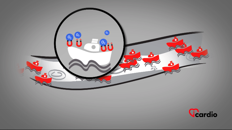
Oxygen supply depends on the number of red blood cells in the blood. These are the “little boats” responsible for transporting oxygen in the blood.

Reliable Pipes Are Essential
The oxygen supply also depends on the state of the piping that irrigates the heart, i.e., the coronary arteriesThe two coronary arteries, the right and the left, form the blood network that supplies the heart with oxygen and nutrients. They are located directly on the surface of the heart and branch into smaller vessels that.
Those Pipes Are Made Up of 3 Layers
The coronary arteriesThe two coronary arteries, the right and the left, form the blood network that supplies the heart with oxygen and nutrients. They are located directly on the surface of the heart and branch into smaller vessels that, like all the other arteries in the human body, are made up of 3 layers.
Each plays a specific role, but the central layer is particularly important in the development of cholesterolCholesterol is essential for the proper functioning of the human body, but it can also have harmful effects if present in excess. >> plaques.


Internal layer: intima
The internal layer, the intima, is like a thin coating of Teflon. It is responsible for the integrity of the vessel and protects it from the formation of bloodBlood is composed of red blood cells, white blood cells, platelets, and plasma. Red blood cells are responsible for transporting oxygen and carbon dioxide. White blood cells make up our immune defense system. Platelets contribute to blood clots.
The intima naturally releases substances used to lubricate and dilate the vessel when necessary, preventing harmful matters from adhering to its wall.
One of the secreted substances can be compared to nitroglycerin which some patients use to soothe chest pain caused by angina. The intima thus helps to keep the vessel well enlarged, in case of need.
Middle layer: media
The media, in the center, is thicker and made up of smooth muscle cells. It gives the artery the ability to spasm or dilate.
The nitroglycerin some people use for angina acts on this layer. It induces vessel relaxation. The vessel becomes larger, circulation improves and angina is relieved.
The media layer is also where cholesterolCholesterol is essential for the proper functioning of the human body, but it can also have harmful effects if present in excess. >> plaques settle, giving arteries a yellowish appearance, as seen from the outside.
External layer: adventitia
The adventitia is the external coating. It is the most resistant part of the artery and offers a protective effect to the vessel.


