The first coronary angiography was performed in 1945.
This examination allows the professional to visualize the arteries of the heart, known as coronaries. It enables the detection of obstructions, such as atheromatous plaques, in these vessels.
The Doctor Makes the Request
The patient does not need to schedule an appointment for this exam.
The cardiologist typically requests it after meeting with the patient in a context suggesting possible coronary arteryThe two coronary arteries, the right and the left, form the blood network that supplies the heart with oxygen and nutrients. They are located directly on the surface of the heart and branch into smaller vessels that blockage. In this context, the request is sent to a specialized center and is done on an outpatient basis, meaning without hospitalization.
Sometimes it's Urgent
Certain circumstances require emergency coronary angiography, such as a myocardial infarction. Other situations, like hospitalization due to unstable angina or other cardiovascular conditions, may necessitate this examination on a semi-urgent basis.
Before the Exam
Often, the hospital calls the patient for a pre-admission appointment. When the patient arrives at the hospital, bloodBlood is composed of red blood cells, white blood cells, platelets, and plasma. Red blood cells are responsible for transporting oxygen and carbon dioxide. White blood cells make up our immune defense system. Platelets contribute to blood tests and an electrocardiogram are performed.
The examination is explained to the patient, and any questions they might have are answered at that time. Generally, this pre-admission takes place a few weeks before the procedure.
Consent Form
After being informed of the risks associated with this examination, the patient must sign a consent form. These risks are detailed later in this text.
At this stage, the physician considers that the benefits of the examination outweigh the risks.
The Day of the Exam
Generally, the patient is hospitalized for only one day for this exam. They must be fasting and should not take any medications from midnight the night before, unless otherwise indicated during the pre-admission.
Upon arrival, the patient is asked to remove all clothing and put on a hospital gown. A nurse then inserts one or two catheters into a vein, through which intravenous fluids and medications can be administered if necessary before the exam.
The hair on the right or left wrist and both groin areas will be shaved to allow for the insertion of catheters into the artery chosen by the cardiologist.
Preparation in the Exam Room
At the appropriate time, the patient is brought into the exam room on a stretcher.
During the exam, the nurses and the doctor take the same precautions as in an operating room to prevent bacteria from entering the patient’s body. The doctor and the team are dressed in blue or green scrubs and wear surgical masks and caps.
The room is kept cool, even cold, to ensure the proper functioning of the radiology equipment. A sterile blanket covers the patient during the exam to minimize discomfort from the room’s temperature and to maintain a sterile environment for the healthcare professionals.
The preparation for the exam is often longer than the exam itself. There are two arteries that ensure circulation in the hand, presenting a particularity. These two arteries have connections; if one gets blocked, the other can compensate. A hand test is often performed to check the reciprocity between the two arteries, or in other words, to verify if the connections are functioning correctly.
In some cases, the radial artery (wrist artery) may be unusable; the femoral artery (groin artery) will then be used to access the heart. The advantages of using the radial artery are the reduced risk of bleeding and the possibility of standing up one to two hours after the exam.
An iodine-based liquid is injected into the coronary arteriesThe two coronary arteries, the right and the left, form the blood network that supplies the heart with oxygen and nutrients. They are located directly on the surface of the heart and branch into smaller vessels that to make them visible on the screen using X-rays. Medical staff wear protective clothing to shield themselves from daily exposure to X-rays.
After preparing the equipment for the procedure, the nurse disinfects the groin and right or left wrist, depending on availability and the exam requirements. Once these areas are dry, she places a sterile blanket over the patient and connects the devices that will be used during the exam. The patient should limit their movements as much as possible from this point on.
During the Exam
The doctor introduces themselves, and the exam begins.
The nurse administers medications to the patient to help them relax and reduce pain during the puncture.
The doctor selects the artery through which the catheters will be inserted.
A Needle Prick
The doctor administers a local anesthetic to minimize pain.
In this case, the wrist artery is used.
A small incision is made, and using a hollow needle, the chosen artery is punctured.
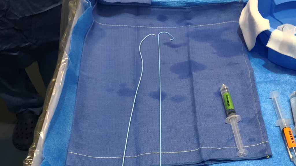
Placement of the Entry Port
A plastic tube, called an introducer, is placed into the artery and keeps this entry accessible for the duration of the exam. The rest of the procedure is painless. The same process is followed when the groin artery is used.
Through this entry port, a long metal wire and a catheter, a long plastic tube, are inserted to reach the coronary arteriesThe two coronary arteries, the right and the left, form the blood network that supplies the heart with oxygen and nutrients. They are located directly on the surface of the heart and branch into smaller vessels that. Once in place, the wire is removed, and the catheter is maneuvered to access the coronary arteriesThe two coronary arteries, the right and the left, form the blood network that supplies the heart with oxygen and nutrients. They are located directly on the surface of the heart and branch into smaller vessels that.
Image Creation
When the catheter is in place, an iodine-based dye is injected.
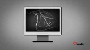
This allows the pathway of the substance inside the coronary arteriesThe two coronary arteries, the right and the left, form the blood network that supplies the heart with oxygen and nutrients. They are located directly on the surface of the heart and branch into smaller vessels that to be clearly seen on images captured by the radiological device that moves around the patient.
These images highlight the presence of partial or complete obstructions and provide information on the size of the arteries, their position, and their anatomical features.
The Moment of Intense Warmth
Once all the images of the coronary arteriesThe two coronary arteries, the right and the left, form the blood network that supplies the heart with oxygen and nutrients. They are located directly on the surface of the heart and branch into smaller vessels that are taken, the exam is almost complete.
Most of the time, the cardiologist will take an additional image to visualize the function of the left ventricle, one of the four heart chambers. This final image helps assess the contraction and performance of the patient’s heart.
During this injection, the patient feels a sudden intense warmth spreading from the head to the hips, which can create the sensation of urinating. This feeling is normal and is caused by the iodine contrast dye injected.
After the Exam
The catheters are removed. A compression bracelet is placed on the patient’s wrist to prevent bleeding while the small hole in the artery closes. This bracelet is gradually loosened over the following hours.
If the procedure is done through the femoral artery, a compression bandage is applied to prevent and control bleeding. The leg must remain stable for four to six hours. Most of the time, an arterial closure device can also be used to seal the small hole in the artery instead of manual compression.
The cardiologist who performed the exam provides the patient with the results and can advise on the next steps. The discovery of obstructions leads to a variety of treatments, ranging from medication prescriptions to angioplasty (unblocking with a balloon and stent) or bypass surgery.
At-Home Recommendations
After a coronary angiography, there are few precautions to take, but it is important to follow these recommendations for the first 4 days:
Do not soak the small wound at the entry site of the exam, whether at the groin or wrist. It is advised to avoid swimming pools and bodies of water. However, it is possible to shower.
If the procedure was done via the wrist, avoid repeated and intense movements of that hand.
For those whose groin artery was used, it is recommended to move the leg every hour if seated for an extended period.
The Results are sent to Your Doctor
The results are communicated to the doctor who asked for the exam.
You may ask for a Copy for Another Doctor
You may ask for a copy of your results to be sent to another doctor. You simply have to give the name and contact information to the personnel. You may ask at any moment.
Procedure Risks
Coronary angiography is a very safe examination, but it carries risks like any other medical procedure performed inside the human body.
The risk of death related to this examination is estimated to be less than 1 in 10,000.
Other major complication risks, such as stroke, heart attack, kidney problems, or the need for urgent surgery, are estimated to be less than 1 in 1,000.
The risk of minor complications, such as bleeding or a hematoma (an accumulation of bloodBlood is composed of red blood cells, white blood cells, platelets, and plasma. Red blood cells are responsible for transporting oxygen and carbon dioxide. White blood cells make up our immune defense system. Platelets contribute to blood), is estimated to be around 1%.