Stress Cardiac Magnetic Resonance Imaging (Cardiac MRI)
Stress cardiac MRI is a more comprehensive version of the examination performed at rest.
It evaluates the structure and function of the heart when it is stimulated to simulate physical effort.
The coronary arteriesThe two coronary arteries, the right and the left, form the blood network that supplies the heart with oxygen and nutrients. They are located directly on the surface of the heart and branch into smaller vessels that are not visualized directly. Instead, the test observes the effects of reduced bloodBlood is composed of red blood cells, white blood cells, platelets, and plasma. Red blood cells are responsible for transporting oxygen and carbon dioxide. White blood cells make up our immune defense system. Platelets contribute to blood supply on the heart muscle.
To do this, images obtained during stress are compared with those taken at rest.
If a region of the heart contracts normally at rest but shows reduced contraction when demand increases, this may suggest that it is not receiving sufficient bloodBlood is composed of red blood cells, white blood cells, platelets, and plasma. Red blood cells are responsible for transporting oxygen and carbon dioxide. White blood cells make up our immune defense system. Platelets contribute to blood flow during exertion. This situation may be related to a narrowing of a coronary arteryThe two coronary arteries, the right and the left, form the blood network that supplies the heart with oxygen and nutrients. They are located directly on the surface of the heart and branch into smaller vessels that.
Like resting cardiac MRI, this examination also analyzes the quality of heart tissue and can detect scarring, inflammation, or other abnormalities.
Stress cardiac MRI does not use ionizing radiation. Unlike X-rays, CT scans, or certain nuclear medicine tests, it relies on a powerful magnetic field (1.5 or 3 Tesla), radiofrequency waves, and an advanced computer system to produce detailed images.
It is therefore recognized as a safe examination when performed according to established protocols.
How Is “Stress” Induced?
Unlike a traditional exercise stress test performed on a treadmill, MRI requires the body to remain still during image acquisition. For technical and logistical reasons, physical exercise cannot be used.
Instead, “stress” is induced by administering a medication intravenously.
Depending on the clinical situation, different substances may be used (such as dipyridamole, adenosine, or dobutamine). These medications temporarily increase the workload of the heart or alter coronary bloodBlood is composed of red blood cells, white blood cells, platelets, and plasma. Red blood cells are responsible for transporting oxygen and carbon dioxide. White blood cells make up our immune defense system. Platelets contribute to blood flow, thereby reproducing the effects of physical exertion.
This allows physicians to observe how the heart muscle responds when its oxygen demand increases.
Why Is It Important?
At rest, some coronary arteryThe two coronary arteries, the right and the left, form the blood network that supplies the heart with oxygen and nutrients. They are located directly on the surface of the heart and branch into smaller vessels that narrowings may go unnoticed because the heart is not working at full capacity.
When the heart is stimulated, a region receiving insufficient bloodBlood is composed of red blood cells, white blood cells, platelets, and plasma. Red blood cells are responsible for transporting oxygen and carbon dioxide. White blood cells make up our immune defense system. Platelets contribute to blood flow may show reduced contraction. This condition is referred to as ischemia.
The information obtained helps to:
confirm or rule out the presence of coronary arteryThe two coronary arteries, the right and the left, form the blood network that supplies the heart with oxygen and nutrients. They are located directly on the surface of the heart and branch into smaller vessels that disease
assess its significance
guide management, whether medical or interventional (angioplasty or surgery)
A Complement to Stress Echocardiography
Stress cardiac MRI can be particularly useful when stress echocardiography does not provide sufficiently precise images.
It then offers a more detailed assessment of heart function.
Scheduling the Appointment
An appointment is required.
In many cases, the referring physician arranges the test. The medical office then communicates the scheduled date and time.
Duration
The examination generally lasts between 60 and 65 minutes.
Special Preparation
Specific instructions are provided before the appointment.
Fasting is required on the day of the examination.
Certain medications must be temporarily discontinued before the test. The physician will specify which ones and when to stop them.
Similarly, coffee, tea, and caffeinated beverages should be avoided for 24 hours prior to the examination, as these substances may interfere with the medication used to simulate exercise.
On the Day of the Appointment
A safety questionnaire must be completed before the procedure. It is designed to verify:
the presence of claustrophobia or discomfort in enclosed spaces
the presence of a cardiac pacemaker
any allergies, particularly to gadolinium
pregnancy
kidney function (a bloodBlood is composed of red blood cells, white blood cells, platelets, and plasma. Red blood cells are responsible for transporting oxygen and carbon dioxide. White blood cells make up our immune defense system. Platelets contribute to blood test may be required if necessary)
the presence of implants or metallic fragments, especially in the brain or eyes
in the context of a stress examination, the presence of asthma
Changing for the Examination
You will be asked to remove clothing from the upper body and wear a hospital gown.
Metal Objects
No metallic objects may be kept on the body (coins, jewelry, belt, etc.). The procedure is similar to airport security measures.
Final Safety Checks
Before entering the examination room, the technologist reviews the questionnaire and confirms all safety instructions.
Placement of Intravenous Lines
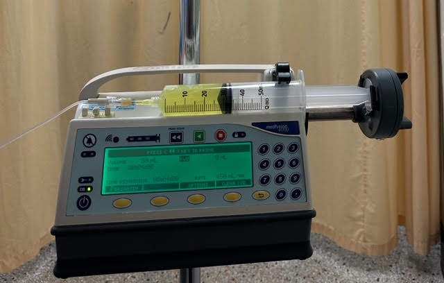
One or, depending on the protocol, two intravenous catheters are inserted into the forearms. These allow administration of the medication used to simulate stress as well as the contrast agent required for image acquisition.
You are then accompanied to the imaging room.
Positioning
The room is well lit and generally maintained at a cool temperature.
Once seated on the movable table, three electrodes are placed on the chest to monitor heart activity throughout the procedure. You may then lie comfortably on your back.
Headphones, a pillow, and a blanket are provided to ensure maximum comfort.
The Procedure
The table gently slides into the machine, which is shaped like a large ring (often compared to a donut).
Only the head and upper part of the body are positioned inside the tunnel. The tunnel is open at both ends, which greatly reduces the feeling of confinement.
No effort is required: the table moves automatically.
Continuous Monitoring
Any discomfort or concern can be communicated at any time.
Once positioning is complete, the technologist moves to the adjacent control room. A glass partition allows continuous observation throughout the examination.
Communication remains constant through the headphones and microphones installed in the room.
A Noisy Machine
The MRI produces intermittent and sometimes loud sounds while images are being acquired.
The headphones help reduce these sounds and allow instructions from the control room to be heard clearly. Between sequences, it is possible to listen to music or the radio, depending on the preference expressed before the examination begins.
The Importance of Breathing Instructions
At certain moments, specific breathing instructions are given.
Carefully following these directions is essential to optimize image quality.
Blood pressure and heart rate
BloodBlood is composed of red blood cells, white blood cells, platelets, and plasma. Red blood cells are responsible for transporting oxygen and carbon dioxide. White blood cells make up our immune defense system. Platelets contribute to blood pressure is checked several times during the procedure.
Heart rhythm is also continuously monitored through the electrodes placed on the chest.
This monitoring ensures that the response to the medication used to simulate stress remains appropriate and safe throughout the examination.
The Stress Phase
In a stress cardiac MRI, the examination begins with the so-called “stress” phase.
A medication is administered intravenously (such as dipyridamole, adenosine, or regadenoson) to reproduce the effects of physical exertion on the heart.
When the medication reaches its maximum effect, a first dose of contrast agent (gadolinium) is injected. The images obtained at this time allow evaluation of how the heart muscle responds when oxygen demand is increased.
The Rest Phase
After a delay of approximately ten minutes, a second injection of contrast is administered during the rest phase.
The images obtained at this stage serve as a comparison.
By comparing the two sets of images, it is possible to identify a region of the heart that functions normally at rest but shows reduced contraction under stress. This difference may suggest insufficient bloodBlood is composed of red blood cells, white blood cells, platelets, and plasma. Red blood cells are responsible for transporting oxygen and carbon dioxide. White blood cells make up our immune defense system. Platelets contribute to blood supply related to a narrowing of a coronary arteryThe two coronary arteries, the right and the left, form the blood network that supplies the heart with oxygen and nutrients. They are located directly on the surface of the heart and branch into smaller vessels that.
Some “rapid” protocols include only a stress phase, which helps shorten the overall duration of the examination.
Delayed Contrast Imaging
The examination continues with images taken several minutes after the gadolinium injection.
These sequences allow assessment of heart muscle quality and detection of possible scarring or other tissue abnormalities.
After the Examination
The catheter is removed at the end of the procedure.
You may then get dressed and leave immediately, unless otherwise instructed.
The results are sent to the physician who ordered the examination. The findings will be interpreted and discussed during the follow-up appointment, along with any recommended next steps.
Results
The results of the examination are sent to the physician who ordered the test.
The findings will be explained and any next steps, if necessary, will be discussed during the follow-up appointment.
Copy for Another Physician
It is also possible to request that a copy of the report be sent to another healthcare professional.
Simply provide the name and contact information of the physician, either before or after the examination.