The Formation of the Ventricles and Major Arteries
Starting from the 4th week, a septum begins to form within the primitive ventricle, giving rise to the interventricular septum. This structure is primarily muscular, but its upper section is completed by a thin fibrous portion known as the membranous septum.
This fibrous portion originates from the partitioning of the arterial bulb, which also gives rise to the pulmonary artery and the aorta, two of the main vessels in the circulatory system.
The Formation of the Ventricles and Major Arteries
As early as the 22nd day of embryonic development, the primitive cardiac tube begins to undergo changes, forming bulges and constrictions.
These rudimentary structures mark the areas that will evolve into the various parts of the heart.
Folding of the Cardiac Tube
Around the 23rd day, uneven growth in the cardiac tube causes a specific folding process. This dynamic reorganization positions the primitive atrium and venous connections behind the primitive ventricle and the arterial bulb.
This folding is crucial for organizing the future cardiac chambers and preparing for the differentiation of the ventricles.
The Formation of the Ventricles and Major Arteries
Around the middle of the 4th week, a septum begins to form within the atrioventricular canal, dividing the heart into two distinct parts: a right side and a left side.
A Wall Divides the Two Ventricles
This process, known as ventricular septation, starts from the lower part of the ventricles and progresses upward. During this time, the right and left ventricular chambers continue to grow and expand.
The Muscular Septum
The septum initially develops in a muscular form, resembling that of the left ventricle, which naturally acquires greater thickness than its right counterpart.
However, this structure remains incomplete at this stage, leaving an opening known as the interventricular communication (IVC) in the upper region of the ventricles.
This temporary communication, characteristic of fetal cardiac development, persists until approximately the 7th week.
Formation of the Aorta and Pulmonary Artery
In parallel with the partitioning of the atrioventricular canal, a distinct mechanism occurs within the arterial bulb. Unlike the linear separation observed in the atriaThe atria are the two upper chambers of the heart. They act as reservoirs for blood that will fill the ventricles. and ventricles, this process follows a spiral structure.
This mechanism, known as aortopulmonary septation, divides the arterial bulb into two major arteries: the aorta, which will carry oxygen-rich bloodBlood is composed of red blood cells, white blood cells, platelets, and plasma. Red blood cells are responsible for transporting oxygen and carbon dioxide. White blood cells make up our immune defense system. Platelets contribute to blood to the body, and the pulmonary artery, which will transport oxygen-poor bloodBlood is composed of red blood cells, white blood cells, platelets, and plasma. Red blood cells are responsible for transporting oxygen and carbon dioxide. White blood cells make up our immune defense system. Platelets contribute to blood to the lungs.
This spiral formation is crucial for ensuring the efficient separation of systemic and pulmonary circulations in the mature heart.
Closure of the Opening Between the Two Ventricles
The persistent opening at the top of the muscular septum between the two ventricles closes with the incorporation of a portion of the arterial bulb. This structure completes the separation of the two ventricular cavities.
The Fibrous Septum Unlike the muscular septum, which constitutes the majority of the interventricular partition, this new portion is composed of fibrous tissue, similar to the adventitia—the outer layer of bloodBlood is composed of red blood cells, white blood cells, platelets, and plasma. Red blood cells are responsible for transporting oxygen and carbon dioxide. White blood cells make up our immune defense system. Platelets contribute to blood vessels.
This fibrous segment plays a crucial role in sealing the communication between the ventricles and ensuring a definitive separation of bloodBlood is composed of red blood cells, white blood cells, platelets, and plasma. Red blood cells are responsible for transporting oxygen and carbon dioxide. White blood cells make up our immune defense system. Platelets contribute to blood flows directed to the aorta and the pulmonary artery.
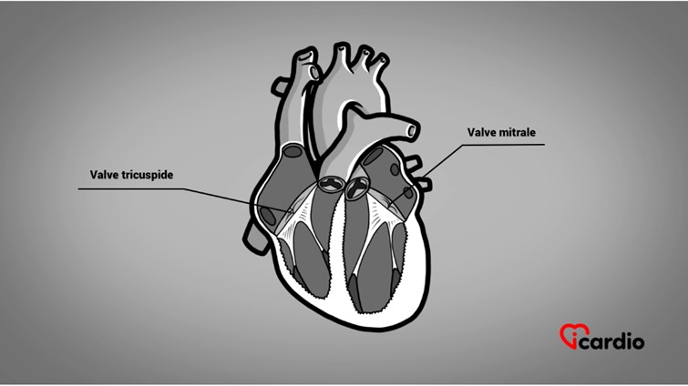
Formation of the Mitral and Tricuspid Apparatus
The ventricles continue their internal development as the spongy muscle bundles undergo gradual reorganization. Some thin out, while others evolve into essential structures: the papillary muscles and chordae tendineae.
These chordae, connected to the papillary muscles, play a crucial role in maintaining the position of the atrioventricular valve leaflets, preventing bloodBlood is composed of red blood cells, white blood cells, platelets, and plasma. Red blood cells are responsible for transporting oxygen and carbon dioxide. White blood cells make up our immune defense system. Platelets contribute to blood reflux during heart contraction.
They include:
The tricuspid valve, made up of three leaflets, located on the right side, regulates the passage between the right atrium and the right ventricle.
The mitral valve, with two leaflets, situated on the left side, controls bloodBlood is composed of red blood cells, white blood cells, platelets, and plasma. Red blood cells are responsible for transporting oxygen and carbon dioxide. White blood cells make up our immune defense system. Platelets contribute to blood flow between the left atrium and the left ventricle.
This precise organization ensures unidirectional bloodBlood is composed of red blood cells, white blood cells, platelets, and plasma. Red blood cells are responsible for transporting oxygen and carbon dioxide. White blood cells make up our immune defense system. Platelets contribute to blood flow, which is essential for the heart’s proper functioning.