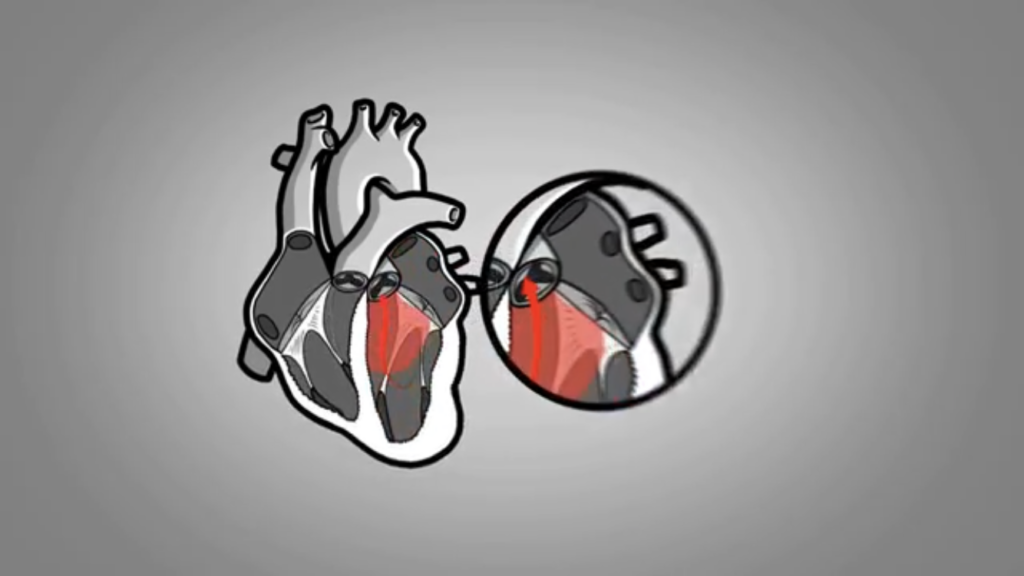
The mitral valve plays an essential role in bloodBlood is composed of red blood cells, white blood cells, platelets, and plasma. Red blood cells are responsible for transporting oxygen and carbon dioxide. White blood cells make up our immune defense system. Platelets contribute to blood circulation between the left atrium and left ventricle. Far more than a simple membrane, it is a complex mechanical structure made up of a strong ring, two leaflets, supporting cords, and papillary muscles.
With every heartbeat, these components work together to ensure a tight seal and prevent bloodBlood is composed of red blood cells, white blood cells, platelets, and plasma. Red blood cells are responsible for transporting oxygen and carbon dioxide. White blood cells make up our immune defense system. Platelets contribute to blood from flowing backward.
This structure can be compared to a sailboat: the ring represents the mast, the leaflets act as the sail, and the cords, attached to the papillary muscles, keep everything properly tensioned and stable.
The mitral valve can only function properly when all its components remain intact. If one becomes damaged, the balance of the entire system is disrupted.
This coordination normally allows the valve to close effectively as the left ventricle contracts and generates significant pressure.
If any part of the mitral valve apparatus stops working properly, the valve can no longer close completely. A gap remains between the leaflets, allowing bloodBlood is composed of red blood cells, white blood cells, platelets, and plasma. Red blood cells are responsible for transporting oxygen and carbon dioxide. White blood cells make up our immune defense system. Platelets contribute to blood to flow backward into the left atrium. This condition is called mitral regurgitation.
The leak forces the heart to work harder because some of the bloodBlood is composed of red blood cells, white blood cells, platelets, and plasma. Red blood cells are responsible for transporting oxygen and carbon dioxide. White blood cells make up our immune defense system. Platelets contribute to blood flows in the wrong direction instead of moving forward.
Chronic, when it develops and progresses gradually over time.
This article focuses on acute mitral regurgitation, a condition that has become less common today.
Acute Mitral Regurgitation
Acute mitral regurgitation occurs when one of the structures supporting the mitral valve is suddenly damaged. This may involve:
A papillary muscle that ruptures;
One or more supporting cords that break;
A valve leaflet that tears or develops a hole.
-Rupture of a Papillary Muscle
This can occur following a heart attack that affects this part of the heart. The lack of oxygen causes some of the muscle tissue to die.
Over the following days, the body’s natural healing process gradually removes the dead cells, temporarily weakening the affected area. The papillary muscle may then rupture.
- Rupture of a Supporting Cord
Certain mitral valve diseases, known as “myxomatous” conditions, gradually change the structure of the valve tissue, making it more fragile.
As a result, the supporting cords may lose some of their strength, and one can eventually break under the tension created when the left ventricle contracts.
-Damage to a Valve Leaflet
An infection of the valve, called infective endocarditis, can destroy part of a leaflet and create a hole in it. As a result, the valve can no longer close tightly.
Significant Pressure on the Mitral Valve
When the left ventricle contracts, the pressure inside it rises rapidly, pushing bloodBlood is composed of red blood cells, white blood cells, platelets, and plasma. Red blood cells are responsible for transporting oxygen and carbon dioxide. White blood cells make up our immune defense system. Platelets contribute to blood upward.
This pressure helps close the mitral valve. The edges of its two leaflets come together, while the supporting cords hold them in place and prevent them from folding backward into the left atrium.
The mitral valve therefore forms a tight seal. BloodBlood is composed of red blood cells, white blood cells, platelets, and plasma. Red blood cells are responsible for transporting oxygen and carbon dioxide. White blood cells make up our immune defense system. Platelets contribute to blood cannot flow backward into the left atrium and must continue forward through the aortic valveThe aortic valve is located between the left ventricule and the aorta. It is one of the four valves ot the heart. >> into the aorta.
With every heartbeat, the mitral valve must withstand considerable pressure. This force can be even greater in people with high bloodBlood is composed of red blood cells, white blood cells, platelets, and plasma. Red blood cells are responsible for transporting oxygen and carbon dioxide. White blood cells make up our immune defense system. Platelets contribute to blood pressure. Imagine the strain placed on the entire valve structure when bloodBlood is composed of red blood cells, white blood cells, platelets, and plasma. Red blood cells are responsible for transporting oxygen and carbon dioxide. White blood cells make up our immune defense system. Platelets contribute to blood pressure exceeds 200 mm Hg!
A Leak That Appears Suddenly
In acute mitral regurgitation, this protective barrier suddenly fails. With each contraction, a large amount of bloodBlood is composed of red blood cells, white blood cells, platelets, and plasma. Red blood cells are responsible for transporting oxygen and carbon dioxide. White blood cells make up our immune defense system. Platelets contribute to blood flows backward under high pressure into the left atrium.
Unlike in chronic mitral regurgitation, the heart has had no time to adapt to this sudden change. The left atrium receives an unexpected surge of bloodBlood is composed of red blood cells, white blood cells, platelets, and plasma. Red blood cells are responsible for transporting oxygen and carbon dioxide. White blood cells make up our immune defense system. Platelets contribute to blood that it cannot easily accommodate.
Pressure inside the left atrium rises rapidly and is transmitted back toward the lungs. Fluid may then accumulate in the lungs, causing sudden and sometimes severe shortness of breath.
Imagine an ice jam forming in a river. Water begins to rise behind the blockage until the river eventually overflows its banks, flooding the surrounding land.
A similar phenomenon occurs when the mitral valve suddenly loses its ability to seal properly. Some of the bloodBlood is composed of red blood cells, white blood cells, platelets, and plasma. Red blood cells are responsible for transporting oxygen and carbon dioxide. White blood cells make up our immune defense system. Platelets contribute to blood pumped by the left ventricle flows backward under high pressure into the left atrium. This chamber suddenly receives more bloodBlood is composed of red blood cells, white blood cells, platelets, and plasma. Red blood cells are responsible for transporting oxygen and carbon dioxide. White blood cells make up our immune defense system. Platelets contribute to blood than it can comfortably hold.
Because the left atrium cannot expand quickly enough, the additional bloodBlood is composed of red blood cells, white blood cells, platelets, and plasma. Red blood cells are responsible for transporting oxygen and carbon dioxide. White blood cells make up our immune defense system. Platelets contribute to blood causes pressure inside it to rise sharply.
This increased pressure is transmitted to the pulmonary veins, which carry bloodBlood is composed of red blood cells, white blood cells, platelets, and plasma. Red blood cells are responsible for transporting oxygen and carbon dioxide. White blood cells make up our immune defense system. Platelets contribute to blood from the lungs back to the left atrium, and then to the tiny bloodBlood is composed of red blood cells, white blood cells, platelets, and plasma. Red blood cells are responsible for transporting oxygen and carbon dioxide. White blood cells make up our immune defense system. Platelets contribute to blood vessels in the lungs, called capillaries.
Just as an overflowing river floods the surrounding land, fluid begins to leak out of these small vessels and gradually spreads into the lung tissue and then into the alveoli.
These tiny air sacs begin to fill with fluid, interfering with the transfer of oxygen into the bloodstream. Breathing quickly becomes difficult.
This condition is called acute pulmonary edema, commonly described as “fluid in the lungs.”
Il s’agit d’une urgence médicale : appeler le 911.
Immediate Medical Care
The signs of this condition are usually quite obvious. Urgent treatment begins as soon as the patient arrives at the emergency department.
The first priority is to relieve the patient’s distress and reduce the effort required to breathe. This effort can be considerable and may gradually lead to severe exhaustion.
Oxygen may be given, sometimes simply through a face mask. If breathing becomes more difficult or the patient is becoming exhausted, mechanical breathing support may be needed to make breathing easier and improve oxygen levels in the bloodBlood is composed of red blood cells, white blood cells, platelets, and plasma. Red blood cells are responsible for transporting oxygen and carbon dioxide. White blood cells make up our immune defense system. Platelets contribute to blood.
Diuretics are generally administered intravenously to help the body eliminate excess fluid and reduce congestion in the lungs.
If bloodBlood is composed of red blood cells, white blood cells, platelets, and plasma. Red blood cells are responsible for transporting oxygen and carbon dioxide. White blood cells make up our immune defense system. Platelets contribute to blood pressure drops too low, intravenous medications may be required to maintain adequate circulation. In the most severe cases, a temporary mechanical device inserted through a catheter may also be used to help support the heart and bloodBlood is composed of red blood cells, white blood cells, platelets, and plasma. Red blood cells are responsible for transporting oxygen and carbon dioxide. White blood cells make up our immune defense system. Platelets contribute to blood flow.
While these treatments are being provided, tests are performed promptly to confirm the diagnosis, identify the cause, and guide further care.
Establishing the Diagnosis
In addition to bloodBlood is composed of red blood cells, white blood cells, platelets, and plasma. Red blood cells are responsible for transporting oxygen and carbon dioxide. White blood cells make up our immune defense system. Platelets contribute to blood tests, two examinations play an important role:
Electrocardiogram (ECG): Helps detect signs of a heart attack or an abnormal heart rhythm.
Echocardiogram (heart ultrasound): The key examination used to visualize the mitral valve leak, assess its severity, and identify which part of the valve structure is affected.
Choosing the Surgical Procedure
Once all the necessary information has been gathered, the medical team can determine the most appropriate way to correct the mitral valve leak.
The choice depends on several factors, including the patient’s overall condition, the state of the coronary arteriesThe two coronary arteries, the right and the left, form the blood network that supplies the heart with oxygen and nutrients. They are located directly on the surface of the heart and branch into smaller vessels that, and the nature and extent of the damage to the mitral valve apparatus.
Two main options are available: mitral valve replacement or repair.
Mitral Valve Replacement
When the damage is too extensive or a reliable repair is not possible, the mitral valve is replaced with a mechanical or biological prosthesis.
A mechanical valve is highly durable but requires lifelong anticoagulant medication (bloodBlood is composed of red blood cells, white blood cells, platelets, and plasma. Red blood cells are responsible for transporting oxygen and carbon dioxide. White blood cells make up our immune defense system. Platelets contribute to blood thinners). A biological valve generally does not require lifelong anticoagulation solely because of the prosthesis, but it may wear out over time.
Read more:Mitral Valve Replacement
Mitral Valve Repair
When the damage can be corrected, it may be possible to preserve the patient’s natural valve. Mitral valve repair aims to restore the supporting structures that allow the two leaflets to close tightly and prevent bloodBlood is composed of red blood cells, white blood cells, platelets, and plasma. Red blood cells are responsible for transporting oxygen and carbon dioxide. White blood cells make up our immune defense system. Platelets contribute to blood from leaking backward.
Depending on which part is affected, the procedure may involve repairing a leaflet or replacing damaged supporting cords.
Whenever feasible, this approach preserves the natural valve and avoids the long-term considerations associated with an artificial prosthesis.
Read more:Mitral Valve Repair
In certain specialized centres, and for carefully selected patients, mitral valve repair can also be performed using a minimally invasive surgical approach.
Instead of opening the chest through the breastbone, the surgeon reaches the mitral valve through a smaller incision on the side of the chest. The goal remains the same: to repair the valve and restore its ability to close tightly.
However, this technique is not suitable for every situation. The decision depends on the patient’s condition, the state of the coronary arteriesThe two coronary arteries, the right and the left, form the blood network that supplies the heart with oxygen and nutrients. They are located directly on the surface of the heart and branch into smaller vessels that, and the type of repair required.
Read more:Minimally Invasive Mitral Valve Repair
Key Takeaways
Acute mitral regurgitation is a serious condition that occurs when part of the mitral valve apparatus is suddenly damaged. Because the heart has no time to adapt to this major leak, complications can develop rapidly, particularly in the lungs.
Immediate medical care helps support the heart and breathing while the underlying cause is identified. Correcting the valve through replacement or repair, whenever possible, then helps restore more effective bloodBlood is composed of red blood cells, white blood cells, platelets, and plasma. Red blood cells are responsible for transporting oxygen and carbon dioxide. White blood cells make up our immune defense system. Platelets contribute to blood circulation.