
Chronic Total Occlusion (CTO) of a Coronary Artery: Medical Overview
- Home
- »
- Coronary heart diseases
- »
- Chronic Total Occlusion (CTO) of a Coronary Artery: Medical Overview

A chronic total occlusion (CTO) is a coronary arteryThe two coronary arteries, the right and the left, form the blood network that supplies the heart with oxygen and nutrients. They are located directly on the surface of the heart and branch into smaller vessels that that has been completely obstructed for at least three months.
Despite this blockage, some people experience no symptoms because of the gradual development of small bloodBlood is composed of red blood cells, white blood cells, platelets, and plasma. Red blood cells are responsible for transporting oxygen and carbon dioxide. White blood cells make up our immune defense system. Platelets contribute to blood vessels called collateral arteries. These natural bypass vessels allow the heart muscle to continue receiving part of the oxygen it needs.

In others, the reduced bloodBlood is composed of red blood cells, white blood cells, platelets, and plasma. Red blood cells are responsible for transporting oxygen and carbon dioxide. White blood cells make up our immune defense system. Platelets contribute to blood supply causes chest pain (angina), shortness of breath, or a reduced ability to exercise. Coronary arteryThe two coronary arteries, the right and the left, form the blood network that supplies the heart with oxygen and nutrients. They are located directly on the surface of the heart and branch into smaller vessels that disease, however, may also progress silently. As a result, some chronic total occlusions are discovered incidentally during tests performed for unrelated reasons.
When symptoms persist despite optimal medical therapy, a specialized procedure may be recommended to reopen the artery and improve quality of life.
Also read: Chronic Total Occlusion (CTO) of a Coronary ArteryThe two coronary arteries, the right and the left, form the blood network that supplies the heart with oxygen and nutrients. They are located directly on the surface of the heart and branch into smaller vessels that: A Patient’s Perspective
In most cases, this type of blockage develops gradually over many years as a result of slowly progressive coronary arteryThe two coronary arteries, the right and the left, form the blood network that supplies the heart with oxygen and nutrients. They are located directly on the surface of the heart and branch into smaller vessels that disease. To better understand how an artery can become completely obstructed, it is helpful to briefly review the role of the coronary arteriesThe two coronary arteries, the right and the left, form the blood network that supplies the heart with oxygen and nutrients. They are located directly on the surface of the heart and branch into smaller vessels that and the processes that gradually narrow them before eventually leading to complete blockage.
Oxygen: Essential for the Heart
The heart is a muscle that works continuously, beating day and night without stopping. To function properly, it requires a constant supply of oxygen and nutrients.
This supply is provided by the coronary arteriesThe two coronary arteries, the right and the left, form the blood network that supplies the heart with oxygen and nutrients. They are located directly on the surface of the heart and branch into smaller vessels that, which deliver oxygen-rich bloodBlood is composed of red blood cells, white blood cells, platelets, and plasma. Red blood cells are responsible for transporting oxygen and carbon dioxide. White blood cells make up our immune defense system. Platelets contribute to blood to the heart muscle.

When bloodBlood is composed of red blood cells, white blood cells, platelets, and plasma. Red blood cells are responsible for transporting oxygen and carbon dioxide. White blood cells make up our immune defense system. Platelets contribute to blood flow through a coronary arteryThe two coronary arteries, the right and the left, form the blood network that supplies the heart with oxygen and nutrients. They are located directly on the surface of the heart and branch into smaller vessels that is reduced—or stops completely—the amount of oxygen reaching the heart muscle becomes insufficient. Depending on the severity of the blockage and how gradually it developed, the reduced bloodBlood is composed of red blood cells, white blood cells, platelets, and plasma. Red blood cells are responsible for transporting oxygen and carbon dioxide. White blood cells make up our immune defense system. Platelets contribute to blood supply may cause no noticeable problems or may eventually damage the heart muscle.
The Coronary Arteries
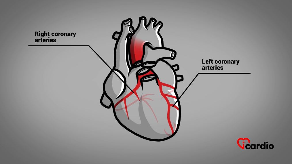
The heart receives its blood supply from two main arteries: the left coronary arteryThe two coronary arteries, the right and the left, form the blood network that supplies the heart with oxygen and nutrients. They are located directly on the surface of the heart and branch into smaller vessels that and the right coronary arteryThe two coronary arteries, the right and the left, form the blood network that supplies the heart with oxygen and nutrients. They are located directly on the surface of the heart and branch into smaller vessels that. Both originate directly from the aorta, the body’s largest artery, which carries oxygen-rich blood from the heart to the rest of the body.
These two arteries branch extensively over the surface of the heart, forming a network that supplies every region of the heart muscle with the oxygen and nutrients it needs to function properly.
Atherosclerotic Plaques
In the vast majority of cases, coronary arteryThe two coronary arteries, the right and the left, form the blood network that supplies the heart with oxygen and nutrients. They are located directly on the surface of the heart and branch into smaller vessels that disease is caused by atherosclerosis, the condition responsible for the formation of atherosclerotic plaques inside the arteries.
Atherosclerosis begins with the buildup of cholesterolCholesterol is essential for the proper functioning of the human body, but it can also have harmful effects if present in excess. >> within the inner lining of the arterial wall. This accumulation triggers an inflammatory response. Cells called macrophages, whose role is to remove waste and harmful substances from the body, absorb the cholesterolCholesterol is essential for the proper functioning of the human body, but it can also have harmful effects if present in excess. >>. As they become filled with fat, they eventually become trapped within the artery wall. When they die, they release their contents, further fueling the inflammatory process.
The body attempts to repair this damaged area by forming a layer of fibrous tissue, similar to a scar, over the plaque. This protective layer helps stabilize the plaque. Over time, calcium deposits may also accumulate, making the plaque harder and less flexible.
Also read: Atherosclerotic Plaque

A Disease That Develops Gradually
Atherosclerosis usually progresses over many years, often without causing any symptoms. Some plaques remain stable, while others continue to grow, gradually narrowing an artery and, in some cases, eventually blocking it completely.
A chronic total occlusion (CTO) may result from:
- the gradual progression of a narrowing until the artery becomes completely blocked;
- a previous heart attack that leaves a coronary arteryThe two coronary arteries, the right and the left, form the blood network that supplies the heart with oxygen and nutrients. They are located directly on the surface of the heart and branch into smaller vessels that permanently occluded;
- changes occurring in a coronary arteryThe two coronary arteries, the right and the left, form the blood network that supplies the heart with oxygen and nutrients. They are located directly on the surface of the heart and branch into smaller vessels that that has already been bypassed with coronary arteryThe two coronary arteries, the right and the left, form the blood network that supplies the heart with oxygen and nutrients. They are located directly on the surface of the heart and branch into smaller vessels that bypass graft (CABG) surgery. In these cases, bloodBlood is composed of red blood cells, white blood cells, platelets, and plasma. Red blood cells are responsible for transporting oxygen and carbon dioxide. White blood cells make up our immune defense system. Platelets contribute to blood flow is maintained by the bypass graft while the native artery may eventually close completely.
As long as the bypass graft continues to function well, no additional treatment is usually required. However, if the graft itself narrows or becomes blocked, reopening the native coronary arteryThe two coronary arteries, the right and the left, form the blood network that supplies the heart with oxygen and nutrients. They are located directly on the surface of the heart and branch into smaller vessels that may be considered.
Chronic Total Occlusion
When a coronary arteryThe two coronary arteries, the right and the left, form the blood network that supplies the heart with oxygen and nutrients. They are located directly on the surface of the heart and branch into smaller vessels that remains completely blocked for at least three months, it is referred to as a chronic total occlusion (CTO).
BloodBlood is composed of red blood cells, white blood cells, platelets, and plasma. Red blood cells are responsible for transporting oxygen and carbon dioxide. White blood cells make up our immune defense system. Platelets contribute to blood can no longer flow through the blocked artery. Nevertheless, the portion of the vessel beyond the obstruction may continue to receive blood through neighboring coronary arteriesThe two coronary arteries, the right and the left, form the blood network that supplies the heart with oxygen and nutrients. They are located directly on the surface of the heart and branch into smaller vessels that.

A Natural Detour Around the Blockage
As a coronary arteryThe two coronary arteries, the right and the left, form the blood network that supplies the heart with oxygen and nutrients. They are located directly on the surface of the heart and branch into smaller vessels that gradually becomes blocked, the heart may compensate by developing small bloodBlood is composed of red blood cells, white blood cells, platelets, and plasma. Red blood cells are responsible for transporting oxygen and carbon dioxide. White blood cells make up our immune defense system. Platelets contribute to blood vessels that bypass the obstruction.
These vessels, known as collateral arteries, may arise from the artery before the blockage or from neighboring coronary arteriesThe two coronary arteries, the right and the left, form the blood network that supplies the heart with oxygen and nutrients. They are located directly on the surface of the heart and branch into smaller vessels that. They function much like secondary roads that allow traffic to bypass a closed highway.
The development of collateral circulation varies greatly from one person to another. In some individuals, it is sufficient to maintain an adequate bloodBlood is composed of red blood cells, white blood cells, platelets, and plasma. Red blood cells are responsible for transporting oxygen and carbon dioxide. White blood cells make up our immune defense system. Platelets contribute to blood supply to the heart muscle, particularly at rest. In others, it remains poorly developed and cannot adequately compensate for the blockage.
Why some people develop an extensive collateral network while others develop very little collateral circulation is still not fully understood.
Even when well developed, collateral arteries have limited capacity. They help keep the heart muscle beyond the blockage alive, but they generally cannot replace the normal bloodBlood is composed of red blood cells, white blood cells, platelets, and plasma. Red blood cells are responsible for transporting oxygen and carbon dioxide. White blood cells make up our immune defense system. Platelets contribute to blood flow provided by a healthy coronary arteryThe two coronary arteries, the right and the left, form the blood network that supplies the heart with oxygen and nutrients. They are located directly on the surface of the heart and branch into smaller vessels that.
How Common Is It?
A chronic total occlusion (CTO) is more common than many people realize. It is estimated that about one in five people (20%) with coronary arteryThe two coronary arteries, the right and the left, form the blood network that supplies the heart with oxygen and nutrients. They are located directly on the surface of the heart and branch into smaller vessels that disease has at least one coronary arteryThe two coronary arteries, the right and the left, form the blood network that supplies the heart with oxygen and nutrients. They are located directly on the surface of the heart and branch into smaller vessels that that has been completely blocked for several months.
Among patients who have previously undergone coronary arteryThe two coronary arteries, the right and the left, form the blood network that supplies the heart with oxygen and nutrients. They are located directly on the surface of the heart and branch into smaller vessels that bypass graft (CABG) surgery, this proportion is even higher, affecting nearly one in two (50%).
Despite its frequency, not every CTO requires treatment. In many people, symptoms are well controlled with medication, while in others, the collateral circulation provides enough bloodBlood is composed of red blood cells, white blood cells, platelets, and plasma. Red blood cells are responsible for transporting oxygen and carbon dioxide. White blood cells make up our immune defense system. Platelets contribute to blood flow to meet the heart muscle’s needs, particularly at rest.

Symptoms… or No Symptoms at All
Learning that a coronary arteryThe two coronary arteries, the right and the left, form the blood network that supplies the heart with oxygen and nutrients. They are located directly on the surface of the heart and branch into smaller vessels that is completely blocked often comes as a surprise, especially when no symptoms are present.
Whether symptoms develop—and how severe they become—depends on several factors, including the amount of heart muscle supplied by the blocked artery, the extent of the collateral circulation, and the heart’s oxygen requirements.
Symptoms usually appear when the heart muscle requires more oxygen than can be delivered by the blocked artery and its collateral vessels. This most often occurs during physical activity, emotional stress, or other situations that increase the heart’s workload.
The most common symptoms include:
- chest pain or pressure (angina), usually during physical activity;
- shortness of breath;
- unusual fatigue;
- reduced ability to perform physical activities or everyday tasks.

In some people, particularly older adults and those living with diabetes, symptoms may be subtle or atypical. Conversely, some CTOs remain completely silent and are discovered incidentally during tests performed for unrelated reasons.
When Should Treatment Be Considered?
Contrary to what many people believe, a chronic total occlusion (CTO) does not always require treatment.
In many cases, symptoms can be managed successfully with medication, healthy lifestyle habits, and the heart’s natural collateral circulation. In these situations, regular medical follow-up is often all that is needed.
However, when symptoms persist despite optimal medical therapy—particularly disabling angina or significant shortness of breath—reopening the blocked artery may be recommended.
The main goals of treatment are to:
- relieve symptoms;
- improve quality of life;
- increase exercise capacity;
- restore bloodBlood is composed of red blood cells, white blood cells, platelets, and plasma. Red blood cells are responsible for transporting oxygen and carbon dioxide. White blood cells make up our immune defense system. Platelets contribute to blood flow to the heart muscle.
The decision to proceed with treatment is always individualized. It takes into account several factors, including the severity of symptoms, their impact on daily life, the results of diagnostic tests, the characteristics of the blockage, and the expected benefits and potential risks of the procedure.
The final decision is made jointly by the patient and the healthcare team to determine the approach that offers the greatest potential benefit for each individual.

Once the Decision Has Been Made
Once treatment has been recommended and accepted, the healthcare team coordinates every step leading up to the intervention. The following stages are designed to ensure the procedure is carried out under the safest possible conditions while answering any remaining questions.
Scheduling the Procedure
Patients do not usually need to schedule a chronic total occlusion (CTO) procedure themselves. In most cases, the cardiologist responsible for follow-up sends a request to a specialized center, usually after a coronary angiogram.
The procedure is then scheduled as an elective intervention, typically within a few days to a few weeks, depending on the patient’s clinical condition and the center’s availability.

Pre-Admission Assessment
Depending on the patient’s medical condition and the hospital’s usual practice, a pre-admission visit may be scheduled before the intervention. However, this step is not necessary in every case.
During this visit, blood tests and an electrocardiogram (ECG) may be performed. The healthcare team also reviews current medications and confirms that everything is in place to perform the procedure safely.
This appointment is an opportunity to explain what to expect, answer any remaining questions, and review the instructions to follow before hospitalization.
If a pre-admission visit is planned, patients should bring an up-to-date list of all their medications.
Informed Consent
Before the procedure, an informed consent form must be signed.
This document confirms that the purpose of the procedure, how it is performed, its expected benefits, and its potential risks have all been explained. It is much more than an administrative form—it is an essential step that allows patients to make a free and informed decision.
It is also the best time to ask any remaining questions, clarify uncertainties, or discuss any concerns. Feeling anxious before a cardiac procedure is entirely normal, but patients should never hesitate to ask for additional explanations. The healthcare team is there to provide guidance and answer every question.
Signing the consent form is entirely voluntary, and consent may be withdrawn at any time, even after it has been signed.

What Are the Risks?
Like any medical procedure, treating a chronic total occlusion (CTO) involves certain risks.
For most people, the greatest concern is naturally the risk of death. Fortunately, when the procedure is performed by an experienced CTO team, this risk remains low—approximately 1%. In patients who have previously undergone coronary arteryThe two coronary arteries, the right and the left, form the blood network that supplies the heart with oxygen and nutrients. They are located directly on the surface of the heart and branch into smaller vessels that bypass graft (CABG) surgery or who have particularly complex coronary anatomy, the risk may increase to 1–2%.
Every treatment decision involves balancing the expected benefits against the potential risks. When a cardiologist recommends reopening a CTO, it is because a careful evaluation indicates that the anticipated benefits outweigh the risks associated with the procedure.
The main risks include:
- Arterial perforation (approximately 1%) – A tear in the artery may allow bloodBlood is composed of red blood cells, white blood cells, platelets, and plasma. Red blood cells are responsible for transporting oxygen and carbon dioxide. White blood cells make up our immune defense system. Platelets contribute to blood to collect around the heart (cardiac tamponade). This complication may require emergency drainage with a needle followed by placement of a small drainage catheter.
- Emergency heart surgery (less than 1%) – In rare cases, emergency cardiac surgery may be required if the complication cannot be managed using catheter-based techniques.
- Death (approximately 1%) – This risk may increase to 1–2% in patients who have previously undergone coronary arteryThe two coronary arteries, the right and the left, form the blood network that supplies the heart with oxygen and nutrients. They are located directly on the surface of the heart and branch into smaller vessels that bypass surgery.
- Bleeding (2–3%) – Bleeding most often occurs at the catheter insertion site in the wrist or groin. Most cases are treated without surgery.
- Stroke (less than 0.5%) – Although uncommon, stroke is a recognized risk of any complex coronary intervention.

Some complications are more specific to CTO procedures.
Because these procedures often take longer than a standard coronary angioplasty, exposure to X-rays is greater. In rare cases, this may cause skin irritation or a superficial skin burn. Thanks to modern imaging equipment, this complication has become very uncommon.
The amount of contrast dye—the liquid injected to make the coronary arteriesThe two coronary arteries, the right and the left, form the blood network that supplies the heart with oxygen and nutrients. They are located directly on the surface of the heart and branch into smaller vessels that visible on X-rays—is also greater than during a routine coronary angioplasty. In people whose kidney function is already reduced, this dye may occasionally cause a temporary decline in kidney function. Preventive measures, including intravenous hydration when appropriate, are routinely used to minimize this risk.
The Day of the Procedure
For many patients, this experience is very similar to having a coronary angiogram. The preparation, the setup in the catheterization laboratory, and the overall course of the intervention are much the same.
Also read: Coronary Angiography
Treatment usually requires a one-day hospital stay. In most cases, patients are asked not to eat or drink after midnight the night before, unless they have received specific instructions regarding their medications.
- Preparation
Upon arrival, a nurse welcomes the patient and prepares everything for the intervention. After changing into a hospital gown, one or two intravenous (IV) lines are inserted to administer fluids and medications if needed.
The wrist and both groins are usually prepared because more than one arterial access site may be required.
Patients are encouraged to empty their bladder before being taken to the cardiac catheterization laboratory. If a lengthy intervention is anticipated, a urinary catheter may occasionally be recommended to improve comfort.
- In the Catheterization Laboratory
The patient is positioned on the procedure table while the medical team prepares a sterile field using precautions similar to those employed in an operating room to minimize the risk of infection.
As with a coronary angiogram, local anesthesia is administered at the arterial access sites. Sedative medications are then given to help the patient relax throughout the intervention.
General anesthesia is usually not required. Patients may drift in and out of sleep while remaining able to answer questions and communicate with the medical team.
The catheterization laboratory is kept cool to ensure the proper functioning of the X-ray equipment. Blankets are provided to help maintain comfort.

A More Challenging Procedure
Although many aspects are similar to those of a coronary angiogram or a routine coronary angioplasty, treating a chronic total occlusion (CTO) is considerably more challenging.
In a conventional angioplasty, the cardiologist treats a narrowed artery through which blood is still able to flow.
When the artery is completely blocked, the first challenge is to find—or recreate—a pathway through the occluded segment before normal bloodBlood is composed of red blood cells, white blood cells, platelets, and plasma. Red blood cells are responsible for transporting oxygen and carbon dioxide. White blood cells make up our immune defense system. Platelets contribute to blood flow can be restored.
This is why the procedure requires specialized techniques, advanced equipment, and a carefully planned strategy tailored to each patient’s coronary anatomy.
Imaging: The Key to Success
The success of the procedure depends largely on the quality of the images obtained during the intervention.
By injecting contrast dye simultaneously through different catheters, the cardiologist can visualize the artery on both sides of the blockage. The collateral arteries often help define the exact course of the occluded segment.
These images provide essential information, including:
- the length of the blockage;
- the amount of calcium present;
- the degree of vessel tortuosity (curvature);
- the quality of the collateral circulation.
Together, these findings guide the choice of technique and play a crucial role in maximizing the likelihood of success.
How Is the Blocked Artery Reopened?
Depending on the characteristics of the blockage, two main approaches may be used.
The Antegrade Approach
With the antegrade approach, the guidewire is advanced in the normal direction of bloodBlood is composed of red blood cells, white blood cells, platelets, and plasma. Red blood cells are responsible for transporting oxygen and carbon dioxide. White blood cells make up our immune defense system. Platelets contribute to blood flow, starting at the beginning of the blocked artery.


The Retrograde Approach
With the retrograde approach, the cardiologist advances through the collateral arteries to reach the artery beyond the blockage before working backward through the occluded segment.


The choice of approach depends entirely on the patient’s coronary anatomy and the characteristics of the blockage.
Advanced Equipment
Crossing a completely blocked coronary arteryThe two coronary arteries, the right and the left, form the blood network that supplies the heart with oxygen and nutrients. They are located directly on the surface of the heart and branch into smaller vessels that requires equipment specifically designed for these highly specialized procedures.
The cardiologist uses guidewires that are extraordinarily thin—barely thicker than a human hair—along with specially designed microcatheters.
When the blockage cannot be crossed directly, it may sometimes be necessary to temporarily create a pathway between the different layers of the arterial wall, bypass the obstruction, and then return to the inside of the artery.

Continuous advances in guidewire technology, microcatheters, and imaging systems have dramatically improved the success rates of this treatment over the past decade.
Reopening the Artery
Once the blockage has been crossed, the guidewire serves as a rail that allows small balloons to be advanced into the occluded segment. These balloons are gradually inflated to create a new passage through the obstruction.
Drug-eluting stents—small metal mesh tubes coated with medication—are then deployed to keep the artery open and maintain long-term bloodBlood is composed of red blood cells, white blood cells, platelets, and plasma. Red blood cells are responsible for transporting oxygen and carbon dioxide. White blood cells make up our immune defense system. Platelets contribute to blood flow.

How Long Does the Procedure Take?
Reopening a chronic total occlusion (CTO) generally takes longer than a conventional angioplasty.
The procedure typically lasts between 90 minutes and 4 hours, depending on the complexity of the blockage.
It requires a high level of precision, adaptability, and sometimes several attempts before the blockage can be successfully crossed. Patience, the expertise of the medical team, and the technologies available all play an essential role in achieving a successful outcome.
Throughout the procedure, patients should report any discomfort so that medications can be administered, if necessary, to improve their comfort.
Success Rates
When performed by experienced operators, current techniques are successful in approximately 90% of cases.
Results depend on several factors, including the complexity of the blockage, the anatomy of the coronary arteriesThe two coronary arteries, the right and the left, form the blood network that supplies the heart with oxygen and nutrients. They are located directly on the surface of the heart and branch into smaller vessels that, and the expertise of the interventional team.

What Are the Expected Benefits?
When reopening the blocked artery successfully restores adequate bloodBlood is composed of red blood cells, white blood cells, platelets, and plasma. Red blood cells are responsible for transporting oxygen and carbon dioxide. White blood cells make up our immune defense system. Platelets contribute to blood flow, most patients experience an improvement in symptoms during physical activity.
This usually translates into better exercise tolerance and an improved quality of life.
In some patients, particularly when the heart muscle remains viable, partial recovery of heart function may also occur. In carefully selected cases, restoring blood flow may also have a favorable impact on long-term prognosis.
After the Procedure
Most patients remain in the hospital overnight for routine monitoring.
During the first few days, it is recommended to avoid soaking the catheter insertion sites in water. Baths and swimming are generally discouraged for 3 to 4 days, although showering is permitted.
Physical activity can usually be resumed gradually after a few days, depending on individual recovery. Most patients are able to return to work within the first week, depending on the nature of their occupation. The appropriate timing should always be discussed with the healthcare team.
Driving should also be avoided for 48 hours following the procedure.
If significant pain, bleeding, or an enlarging lump develops at the catheter insertion site, prompt evaluation in an emergency department is recommended.
Preventing Blood Clots Inside the Stent
Following stent implantation, medication is essential to prevent the formation of a blood clot inside the stent—a complication known as stent thrombosis.
Treatment usually consists of dual antiplatelet therapy (DAPT):
- aspirin, which is generally continued long term;
- a second antiplatelet medication, prescribed for at least several months—often up to one year, and sometimes longer depending on the clinical situation.
Strict adherence to this treatment is essential to keep the artery open and minimize the risk of stent thrombosis.
Also read: About Coronary Stents

Never stop taking an antiplatelet medication during the first year after stent implantation without first consulting your cardiologist—even before dental work or any surgical procedure.

Follow-Up Care
Any concerns related to treatment should be discussed with a physician familiar with coronary interventions, ideally a cardiologist.
Over the months following stent implantation, the artery gradually heals and forms a new layer of tissue over the stent. Depending on the patient’s progress, this healing process may eventually allow the second antiplatelet medication to be discontinued—but only on the recommendation of the treating cardiologist.
Conclusion
A chronic total occlusion (CTO) of a coronary arteryThe two coronary arteries, the right and the left, form the blood network that supplies the heart with oxygen and nutrients. They are located directly on the surface of the heart and branch into smaller vessels that is a common condition that may develop silently over many years. In some individuals, collateral arteries partially compensate for the blockage, while in others, symptoms such as angina or shortness of breath gradually appear.
Recent advances have transformed the management of CTOs. Specialized techniques, increasingly sophisticated equipment, advanced imaging technologies, and dedicated teams in specialized centers have significantly improved both the success rates and the safety of these procedures.
When appropriate, reopening the blocked artery can relieve symptoms, improve exercise capacity, and enhance quality of life for most patients. In selected individuals, it may also promote partial recovery of heart function.
The decision to proceed with treatment is always individualized. It is based on a careful assessment of symptoms, the characteristics of the blockage, the expected benefits, and the potential risks of the procedure.
A good understanding of the condition, careful adherence to medical therapy, and regular follow-up with the healthcare team remain the best ways to achieve durable, long-term results.
Related articles


Latest articles