Coronary angiography is an examination that uses a contrast dye visible on X-rays to visualize the arteries of the heart. This technique makes it possible to check for blockages in the coronary arteriesThe two coronary arteries, the right and the left, form the blood network that supplies the heart with oxygen and nutrients. They are located directly on the surface of the heart and branch into smaller vessels that and assess their severity.
Before the Era of Coronary Angiography
Until the late 1950s, no one had ever seen the arteries of the heart in a living person. The only way to discover a coronary blockage was… during an autopsy. In practice, this helped no one: it was impossible to prevent a heart attack or guide treatment.
Cardiologists knew that coronary arteryThe two coronary arteries, the right and the left, form the blood network that supplies the heart with oxygen and nutrients. They are located directly on the surface of the heart and branch into smaller vessels that disease existed — its symptoms had been described for centuries — but they were essentially working in the dark.
No technology allowed them to observe the arteries that supply bloodBlood is composed of red blood cells, white blood cells, platelets, and plasma. Red blood cells are responsible for transporting oxygen and carbon dioxide. White blood cells make up our immune defense system. Platelets contribute to blood to the heart. They could suspect a problem, but never actually see it.
“Do NOT inject contrast into the coronaries!”
At that time, cardiac catheterization was beginning to develop, but several myths persisted. The most widespread belief was that placing a catheter directly into a heart artery — or worse, injecting contrast dye into it — would lead to disastrous consequences.
It was thought this would trigger an immediate and fatal arrhythmia: ventricular fibrillation.
This fear came largely from experiments in animals, where direct injection into a coronary arteryThe two coronary arteries, the right and the left, form the blood network that supplies the heart with oxygen and nutrients. They are located directly on the surface of the heart and branch into smaller vessels that did cause severe arrhythmias. From this, physicians concluded — without ever confirming it in humans — that no person would survive such an injection.
As a result:
➡️ doctors strictly avoided the coronary arteriesThe two coronary arteries, the right and the left, form the blood network that supplies the heart with oxygen and nutrients. They are located directly on the surface of the heart and branch into smaller vessels that, ➡️ they were considered completely off-limits, ➡️ and no one dared insert a catheter into them.
A First Step: Seeing the Coronary Arteries Indirectly
In 1958, radiologist Charles Dotter developed a way to visualize the coronary arteries… without ever touching them. He placed a small catheter with a balloon at its tip at the very beginning of the aorta. By inflating the balloon, he could inject contrast dye only into the aortic root, the point where the coronary arteriesThe two coronary arteries, the right and the left, form the blood network that supplies the heart with oxygen and nutrients. They are located directly on the surface of the heart and branch into smaller vessels that originate.
And then, a small miracle:
➡️ no one died, ➡️ and the openings of the coronary arteriesThe two coronary arteries, the right and the left, form the blood network that supplies the heart with oxygen and nutrients. They are located directly on the surface of the heart and branch into smaller vessels that became visible.
In doing so, he demonstrated that simply allowing contrast dye to flow into the coronaries from the aorta caused no harmful consequences. This was a turning point — the first crack in the myth that any opacification of the coronary arteriesThe two coronary arteries, the right and the left, form the blood network that supplies the heart with oxygen and nutrients. They are located directly on the surface of the heart and branch into smaller vessels that would be fatal.
Without knowing it, Dr. Dotter had just opened the door to true coronary angiography… a breakthrough that would occur a few months later, entirely by accident.
An Accidental Discovery That Changes Everything
A few months later, at the Cleveland Clinic, cardiologist Mason Sones was performing a routine examination: an aortography, a procedure used to visualize the aorta, the largest artery in the human body. This massive vascular “highway” begins directly at the exit of the heart and distributes bloodBlood is composed of red blood cells, white blood cells, platelets, and plasma. Red blood cells are responsible for transporting oxygen and carbon dioxide. White blood cells make up our immune defense system. Platelets contribute to blood to all other arteries.
His patient that day was Abner Darby, 47 years old. The catheter was correctly positioned at the start of the aorta. Sones asked his assistant, Dr. Royson Lewis, to trigger the injection of contrast.
But due to the pressure produced by the automatic injector, the catheter shifted… and entered directly into the right coronary arteryThe two coronary arteries, the right and the left, form the blood network that supplies the heart with oxygen and nutrients. They are located directly on the surface of the heart and branch into smaller vessels that. In one second, 40 mL of contrast — an enormous volume for such a small artery — rushed into the coronary.
Both physicians turned pale. According to everything believed at the time, the patient was expected to die on the table within seconds.
Sones shouted, “Stop! Stop!”, tapped the patient’s chest to stimulate a cardiac reflex, and braced for the inevitable.
But the inevitable… never came.
Mr. Darby experienced a marked but very brief slowing of his heart rate, then his heart gradually returned to a normal rhythm. Just moments earlier, everyone feared the worst… yet the patient was very much alive.
In the lab, disbelief turned into astonishment. What should have been a catastrophe became a historic revelation.
Birth of Selective Coronary Angiography
After this event, Sones understood that it was possible to inject contrast directly into the coronary arteriesThe two coronary arteries, the right and the left, form the blood network that supplies the heart with oxygen and nutrients. They are located directly on the surface of the heart and branch into smaller vessels that safely. He then developed the technique of selective coronary angiography:
➡️ a specific injection into the left coronary arteryThe two coronary arteries, the right and the left, form the blood network that supplies the heart with oxygen and nutrients. They are located directly on the surface of the heart and branch into smaller vessels that, ➡️ then into the right coronary arteryThe two coronary arteries, the right and the left, form the blood network that supplies the heart with oxygen and nutrients. They are located directly on the surface of the heart and branch into smaller vessels that.
For the first time, physicians could clearly see coronary blockages in living patients. The principle used today is… exactly the same.
Finally Understanding Coronary Artery Disease
Before coronary angiography, coronary arteryThe two coronary arteries, the right and the left, form the blood network that supplies the heart with oxygen and nutrients. They are located directly on the surface of the heart and branch into smaller vessels that disease was poorly understood. Its symptoms had been described as far back as the 1700s, but never directly observed. It took another hundred years before pathologists identified blockages in the coronary arteriesThe two coronary arteries, the right and the left, form the blood network that supplies the heart with oxygen and nutrients. They are located directly on the surface of the heart and branch into smaller vessels that.
Beginning in the late 1950s, everything changed:
➡️ doctors could finally see atherosclerotic plaques, ➡️ assess their severity, ➡️ and understand that they caused angina, heart attacks, and sudden death.
This discovery ushered in a new era of treatments: medications, surgery, and eventually angioplasty.
A New Challenge: Atherosclerotic Plaques
When the first images of the coronary arteriesThe two coronary arteries, the right and the left, form the blood network that supplies the heart with oxygen and nutrients. They are located directly on the surface of the heart and branch into smaller vessels that appeared on radiology screens, it was a true revelation. In vessels once assumed to be smooth and clean, physicians suddenly saw bumps, narrowings, and irregular shadows — atherosclerotic plaques formed by cholesterol accumulated over the years.
For the first time, the enemy was visible. And what they discovered was unmistakable:
these plaques can strangle bloodBlood is composed of red blood cells, white blood cells, platelets, and plasma. Red blood cells are responsible for transporting oxygen and carbon dioxide. White blood cells make up our immune defense system. Platelets contribute to blood flow and lead to:
angina,
heart attacks,
and sometimes sudden death.
It was as if someone had finally turned on the lights in a room that had been dark for centuries.
Seeing what was hidden inside these arteries triggered an urgent search for solutions: how to prevent these deposits, how to stabilize them, how to remove them, or bypass them?
This discovery opened the door to a new era — one in which the goal was not only to see the disease… but to treat it.
And What Came Next for Dr. Sones
A true visionary, Dr. Mason Sones worked with engineers to design catheters better suited to this new technique. However, his method used an approach through the brachial artery at the elbow crease, which was more complex.
Ironically, this pioneer of cardiology later died of lung cancer… with a cigarette in hand.
The Arrival of Dr. Melvin Judkins
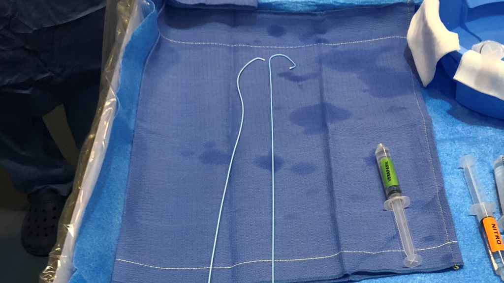
Radiologist Melvin Judkins, trained by Dotter and later sent to work with Sones, quickly realized that the technique could be simplified.
He noticed that, from one person to another, the origin of the right and left coronary arteriesThe two coronary arteries, the right and the left, form the blood network that supplies the heart with oxygen and nutrients. They are located directly on the surface of the heart and branch into smaller vessels that is always located in the same anatomical position.
He therefore developed pre-shaped catheters, advanced through the femoral artery, that were much easier to manipulate.
Thus, the Judkins catheters were born. They made coronary angiography simpler, faster, and safer.
Even today, these left and right Judkins catheters are the ones used daily in cardiac catheterization laboratories around the world.