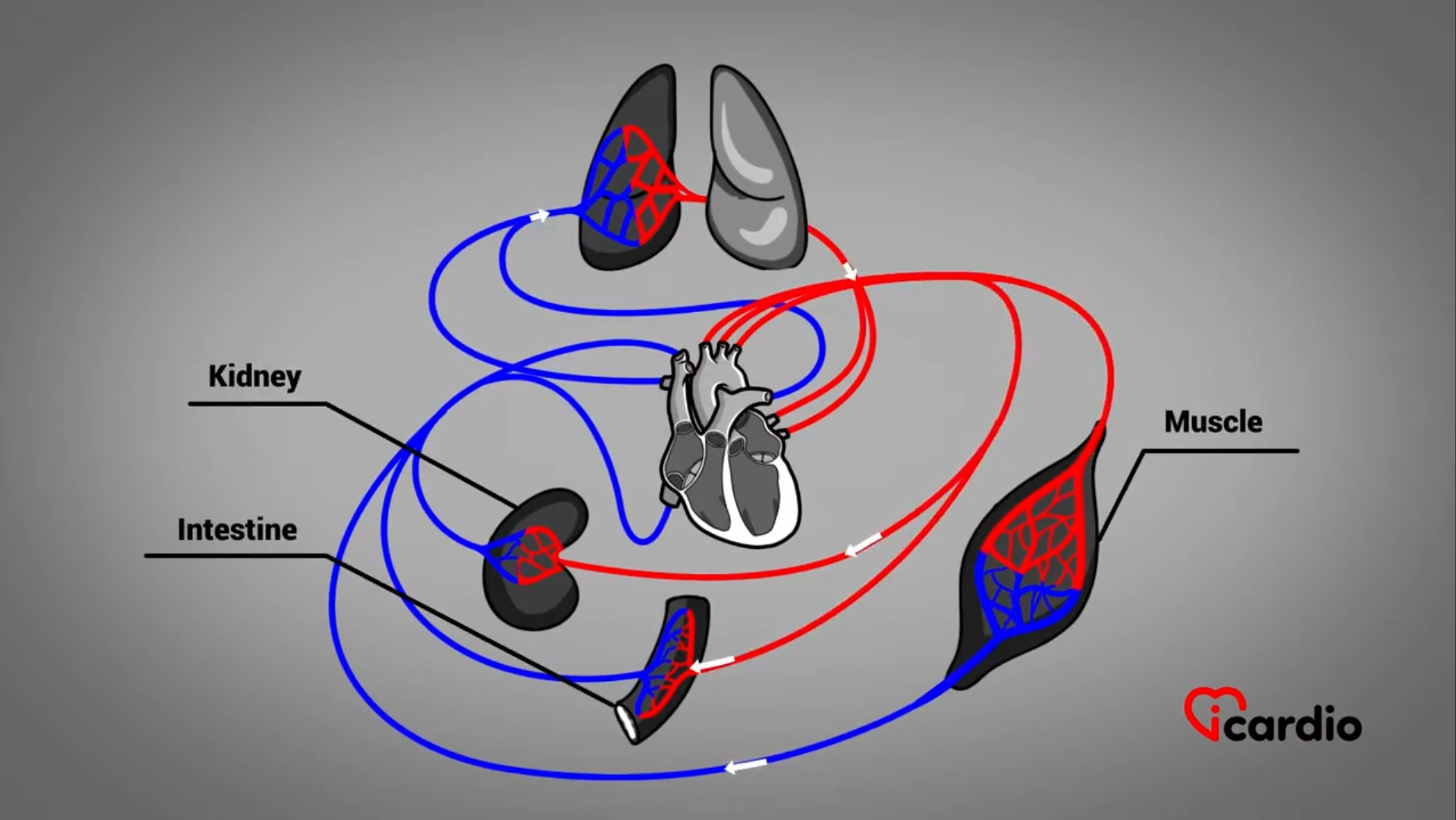
The heart is a muscular organ about the size of a closed fist. It is located in the center of the chest, between the 2 lungs. Il est de la grosseur d’un poing fermé.
The heart is a marvelous, almost indefatigable organ. It enables bloodBlood is composed of red blood cells, white blood cells, platelets, and plasma. Red blood cells are responsible for transporting oxygen and carbon dioxide. White blood cells make up our immune defense system. Platelets contribute to blood to circulate throughout the body.
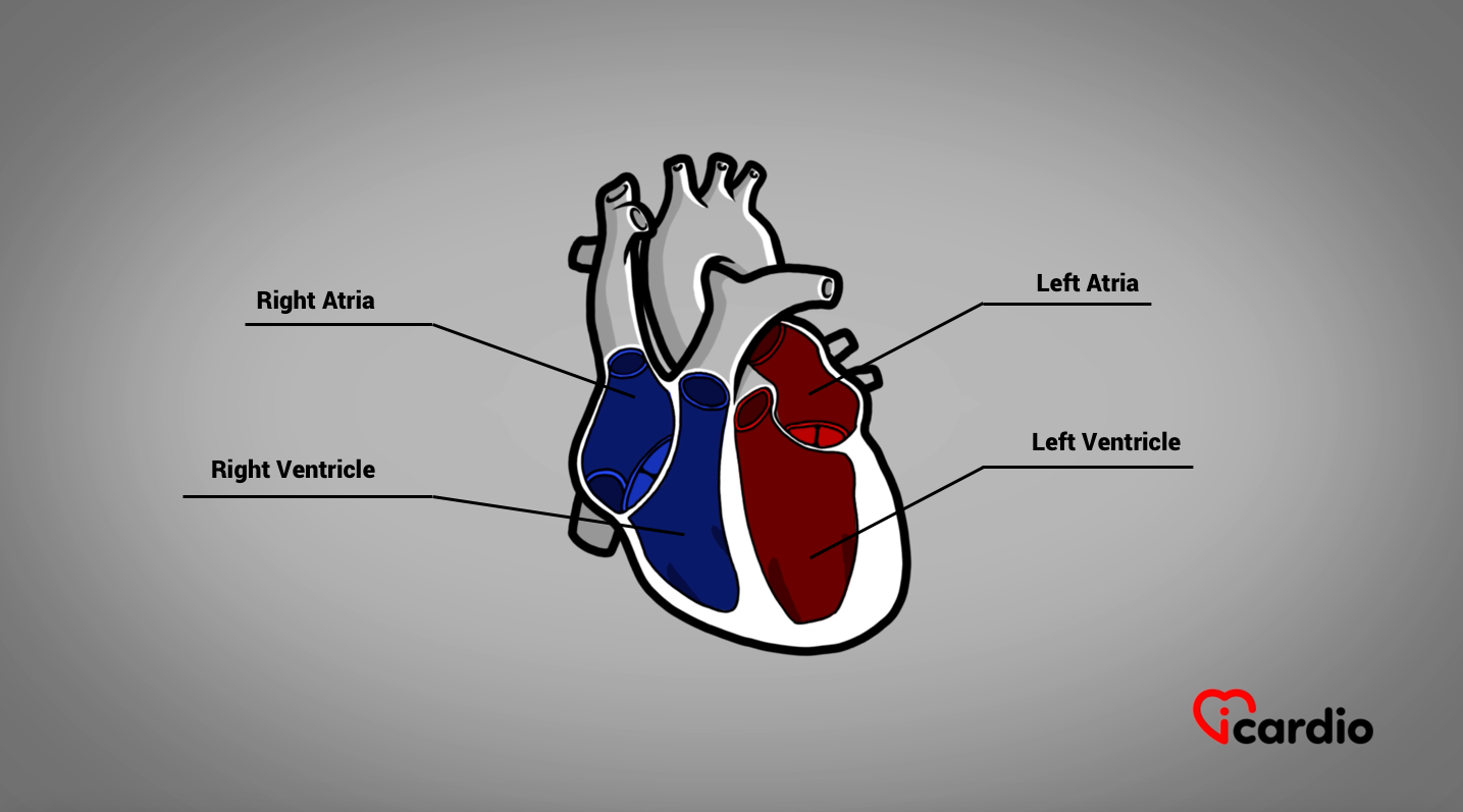
The 4 Chambers of the Heart
The heart is made up of 4 parts called chambers. The 2 upper chambers, the atriaThe atria are the two upper chambers of the heart. They act as reservoirs for blood that will fill the ventricles., are reservoirs for the bloodBlood is composed of red blood cells, white blood cells, platelets, and plasma. Red blood cells are responsible for transporting oxygen and carbon dioxide. White blood cells make up our immune defense system. Platelets contribute to blood that fills the ventricles.
The ventricles are the 2 lower chambers of the heart. They are more muscular than the atriaThe atria are the two upper chambers of the heart. They act as reservoirs for blood that will fill the ventricles.; their function is to propel bloodBlood is composed of red blood cells, white blood cells, platelets, and plasma. Red blood cells are responsible for transporting oxygen and carbon dioxide. White blood cells make up our immune defense system. Platelets contribute to blood to ensure circulation throughout the body.
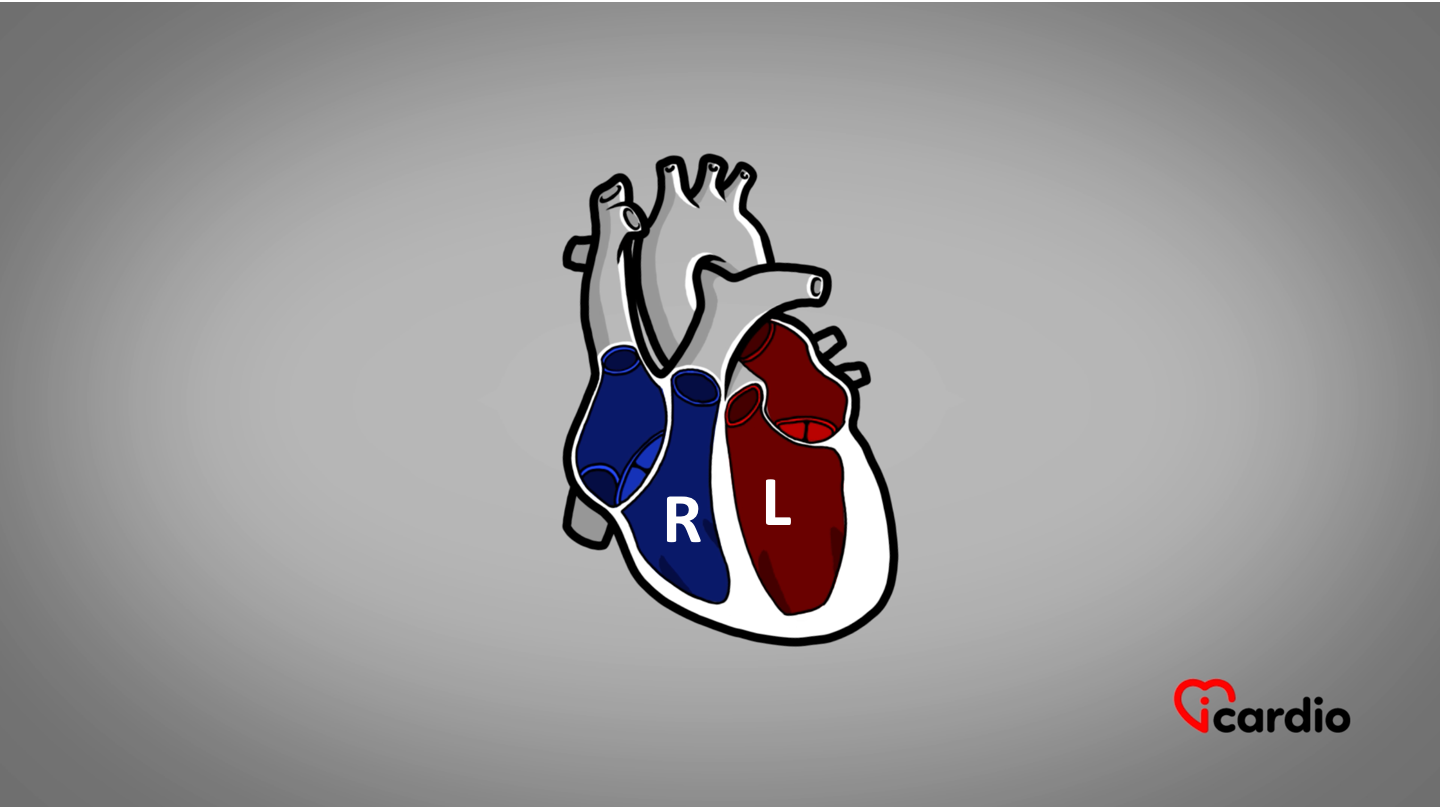
A Right Heart and a Left Heart
The heart can be separated into 2 parts. An internal tissue wall between the chambers divides the right (in blue on the picture) and the left side of the heart (in red), each equipped with an atrium and a ventricle.
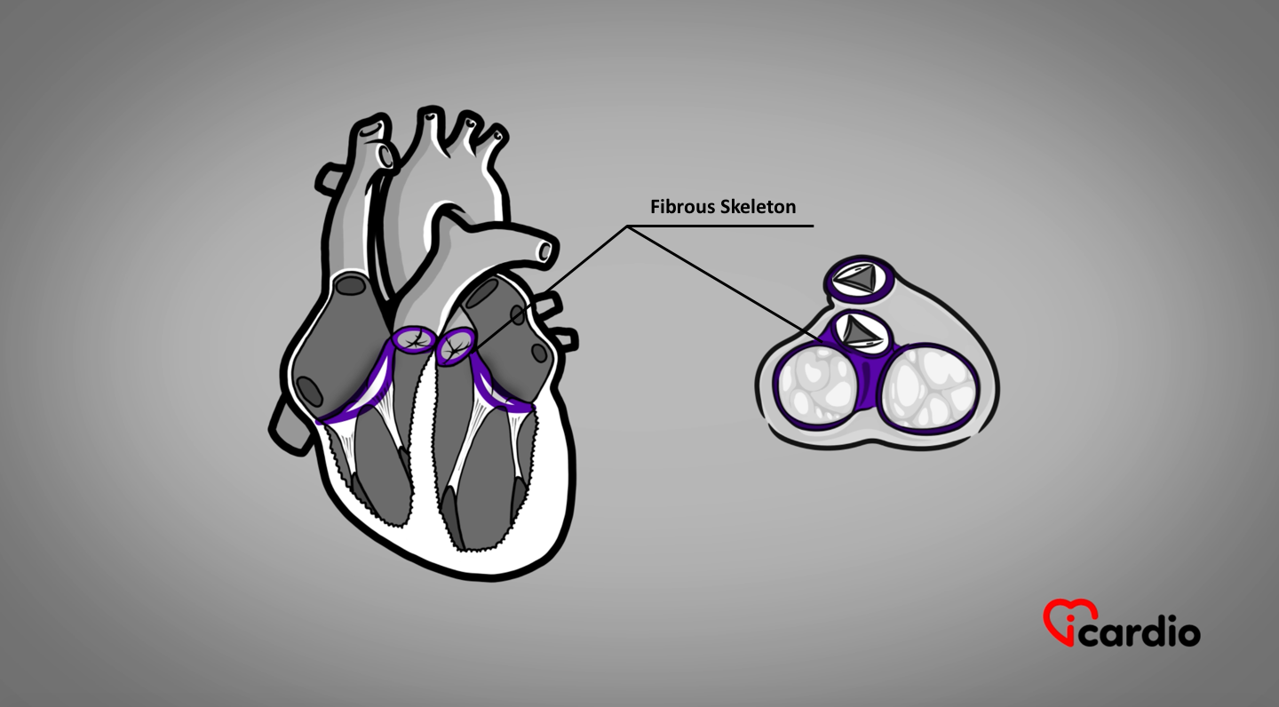
The Heart Is Supported by a Skeleton
The heart rests on a fibrous skeletonLike the human body, the heart has its skeleton. The 4 chambers and valves of the heart are attached to it. In other words, on a rigid structure.
The atriaThe atria are the two upper chambers of the heart. They act as reservoirs for blood that will fill the ventricles. and ventricles are joined by 2 large rings called atrioventricular rings.
The other 2 rings are smaller. They connect the pulmonary artery to the right ventricle and the aorta to the left ventricle.
The 4 Heart Valves
The human heart has 4 valves that control bloodBlood is composed of red blood cells, white blood cells, platelets, and plasma. Red blood cells are responsible for transporting oxygen and carbon dioxide. White blood cells make up our immune defense system. Platelets contribute to blood flow in the right direction… with no possibility of backflow!
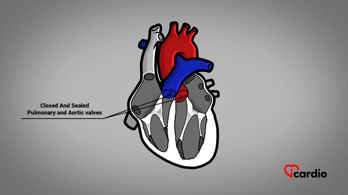
2 of those valves, namely the pulmonary valve and the aortic valveThe aortic valve is located between the left ventricule and the aorta. It is one of the four valves ot the heart. >>, are located at the base of the large vessels, right under the ventricles.
After ejection, the bloodBlood is composed of red blood cells, white blood cells, platelets, and plasma. Red blood cells are responsible for transporting oxygen and carbon dioxide. White blood cells make up our immune defense system. Platelets contribute to blood column tries to flow backward due to the pressure reduction and suction effect caused by the relaxation of the ventricles. The attempt to reverse the flow reopens the cups, blocking the bloodBlood is composed of red blood cells, white blood cells, platelets, and plasma. Red blood cells are responsible for transporting oxygen and carbon dioxide. White blood cells make up our immune defense system. Platelets contribute to blood and sealing the valve.
After ejection, the blood column tries to flow backward due to the pressure reduction and suction effect caused by the relaxation of the ventricles. The attempt to reverse the flow reopens the cups, blocking the blood and sealing the valve.
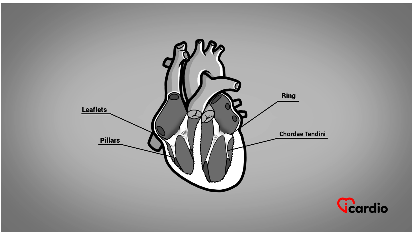
The 2 other heart valves, called atrioventricular valves, are found between the atriaThe atria are the two upper chambers of the heart. They act as reservoirs for blood that will fill the ventricles. and the ventricles.
These valves are hinged to atrioventricular ring-shaped annuli. They are composed of leaflets, cords, and muscular pillars at the ventricular level.
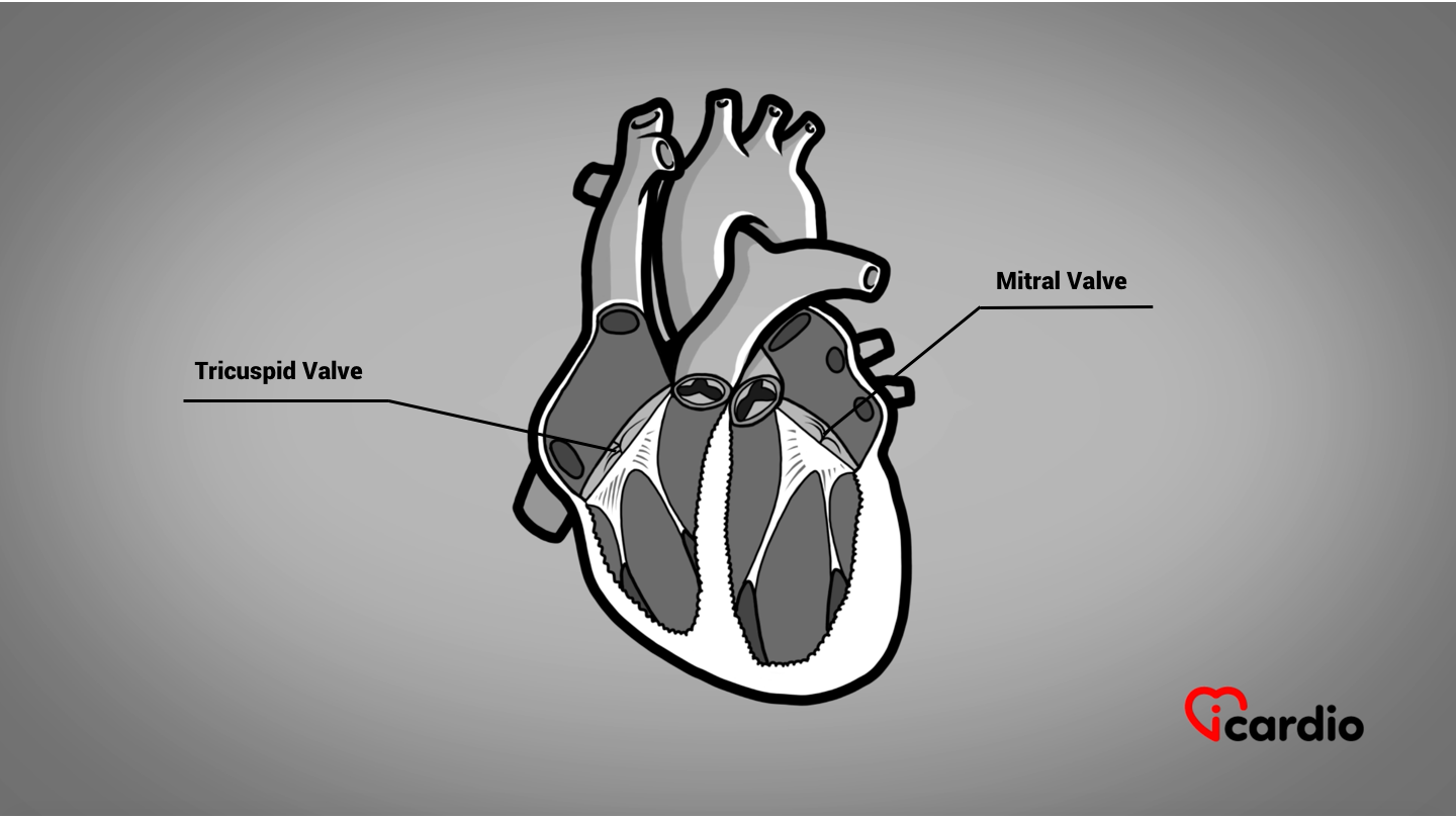
The tricuspid valve is on the right and the mitral valve is on the left.
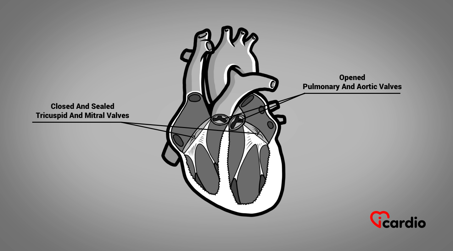
When the ventricles contract, the pressure generated by the muscle pushes the bloodBlood is composed of red blood cells, white blood cells, platelets, and plasma. Red blood cells are responsible for transporting oxygen and carbon dioxide. White blood cells make up our immune defense system. Platelets contribute to blood volume upward, closing the leaflets.
The leaflets are held in place by cords that prevent them from folding back into the atria.
Therefore, the only way out for the bloodBlood is composed of red blood cells, white blood cells, platelets, and plasma. Red blood cells are responsible for transporting oxygen and carbon dioxide. White blood cells make up our immune defense system. Platelets contribute to blood is to go through the pulmonary valve on the right, and the aortic valveThe aortic valve is located between the left ventricule and the aorta. It is one of the four valves ot the heart. >> on the left.
The Lining of the Heart
If we were to rub our hands nonstop for a long time, the continuous friction would produce irritation and eventually inflammation, swelling, and even blisters.
The heart is in constant motion and is inside a protective fluid-filled sac. This substance is a lubricant that allows the heart to slide, without any friction, inside a pouch called the pericardiumThe pericardium is a sac surrounding the heart and containing a lubricating fluid that allows it to glide with each beat without friction..
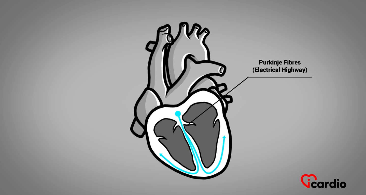
The Heart Has its Own Electrical System
The heart has its very own fascinating electrical system that synchronizes its contraction effectively.
The “band leader” of this system is located at the top of the heart, near the right atrium. It is the heart’s natural pacemaker, initiating all the heartbeats and determining the heart rate.
Transmission between the atria and ventricles is only possible at one point. This location is made up of specialized cells. It controls the speed at which electrical stimulation reaches the ventricles. Beyond a certain frequency of atrial stimulation, the passage is slowed, limiting the number of heartbeats or pulses per unit of time. It is an electrical corridor under surveillance.
As soon as the electricity has crossed the electrical customs barrier, the current is transmitted to the right and left ventricles via 2 branches, i.e. 2 distinctive, fast-conducting electrical wires.
The electrical transmission, originating in the ventricles, enables the heart to contract from bottom to top, projecting bloodBlood is composed of red blood cells, white blood cells, platelets, and plasma. Red blood cells are responsible for transporting oxygen and carbon dioxide. White blood cells make up our immune defense system. Platelets contribute to blood toward the pulmonary and aortic valvesThe aortic valve is located between the left ventricule and the aorta. It is one of the four valves ot the heart. >>.
The Large Vessels Attached to the Heart
The main bloodBlood is composed of red blood cells, white blood cells, platelets, and plasma. Red blood cells are responsible for transporting oxygen and carbon dioxide. White blood cells make up our immune defense system. Platelets contribute to blood vessels running from the heart are the superior and inferior vena cava, the pulmonary artery, the 4 pulmonary veins, and the aorta.
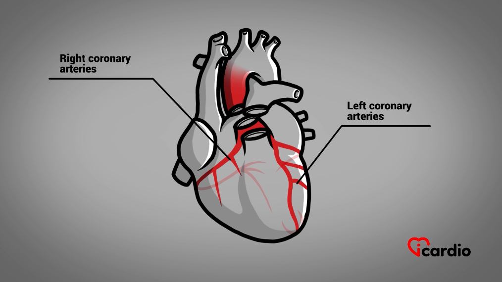
The Arteries that Feed the Heart
The bloodBlood is composed of red blood cells, white blood cells, platelets, and plasma. Red blood cells are responsible for transporting oxygen and carbon dioxide. White blood cells make up our immune defense system. Platelets contribute to blood network supplying the heart is made up of 2 coronary arteriesThe two coronary arteries, the right and the left, form the blood network that supplies the heart with oxygen and nutrients. They are located directly on the surface of the heart and branch into smaller vessels that, one right and one left.