
An abdominal aortic aneurysm is often discovered by chance, without any symptoms. Yet, if it tears or ruptures, the consequences can be serious—even fatal.
The key is to monitor the aneurysm’s evolution regularly in order to determine the right time to intervene.
Careful surveillance and a well-planned surgical decision often prevent an emergency and greatly improve the chances of success.
A Brief Anatomy Lesson
The aorta is the largest artery in the human body. It emerges from the heart, receives bloodBlood is composed of red blood cells, white blood cells, platelets, and plasma. Red blood cells are responsible for transporting oxygen and carbon dioxide. White blood cells make up our immune defense system. Platelets contribute to blood ejected from the left ventricle, and distributes it throughout the body—much like the trunk of a tree sending sap to its branches.
The aorta is divided into three main sections:
- Ascending aorta (A): The initial segment begins at the heart and rises toward the major vessels that supply bloodBlood is composed of red blood cells, white blood cells, platelets, and plasma. Red blood cells are responsible for transporting oxygen and carbon dioxide. White blood cells make up our immune defense system. Platelets contribute to blood to the brain and arms.
- Thoracic aorta (B): From this point, it continues downward through the chest and passes through the diaphragm, the main muscle involved in breathing.
- Abdominal aorta (C): After leaving the thoracic cavity, it travels deep into the abdomen. It eventually divides into two large branches forming an inverted Y—the iliac arteries, which carry bloodBlood is composed of red blood cells, white blood cells, platelets, and plasma. Red blood cells are responsible for transporting oxygen and carbon dioxide. White blood cells make up our immune defense system. Platelets contribute to blood to the legs.

Normal Size
D’un segment à l’autre, l’aorte tend à diminuer de calibre à mesure qu’elle s’éloigne du cœur. Il est normal que ce grand vaisseau subisse une légère dilatation avec l’âge. Toutefois, le diamètre de l’aorte abdominale ne devrait pas excéder 3 centimètres, soit environ un peu plus d’un pouce.
What Is an Aneurysm?
An aneurysm is a localized weakening of the wall of an artery.
The muscular layer that normally gives it strength becomes less resistant. Under the pressure of circulating bloodBlood is composed of red blood cells, white blood cells, platelets, and plasma. Red blood cells are responsible for transporting oxygen and carbon dioxide. White blood cells make up our immune defense system. Platelets contribute to blood, this weakness causes a gradual deformation and dilation at a specific point in the vessel.
It is therefore not the entire aorta that expands, but rather a localized portion, most often situated below the renal arteries, in the lower part of the abdominal aorta.
This phenomenon can be compared to a bulge forming on a garden hose: internal pressure pushes outward where the material is weakest.

When the aorta’s diameter exceeds 3 centimeters, it is considered aneurysmal. Beyond this measurement, the condition is called an abdominal aortic aneurysm—a slow and silent process that carries a real risk of rupture.
Men are affected more often than women, especially after the age of 65. Smoking and high bloodBlood is composed of red blood cells, white blood cells, platelets, and plasma. Red blood cells are responsible for transporting oxygen and carbon dioxide. White blood cells make up our immune defense system. Platelets contribute to blood pressure further increase this likelihood.
Although rupture remains rare, it represents a life-threatening emergency when it occurs. Regular screening and medical follow-up can prevent complications before they become serious.
The most common type of aortic aneurysm is the abdominal aortic aneurysm, but other segments of the aorta can sometimes be affected at the same time.
What Causes This Bulge in the Vessel?
This weakening of the arterial wall is most often caused by the formation of an atherosclerotic plaque.
In rarer cases, certain conditions that directly affect the muscular layer of the vessel can also weaken the wall.
- Genetic disorders, such as Marfan syndrome or other inherited connective tissue diseases, can alter the structure of the aorta.
- Infectious or inflammatory processes can also progressively damage and weaken the arterial wall.
Although these causes are less common, they share the same effect: they make the aorta more vulnerable to dilation and promote the development of an aneurysm.

Atherosclerotic Plaque
It all begins with the deposition of bad cholesterolCholesterol is essential for the proper functioning of the human body, but it can also have harmful effects if present in excess. >> (LDL) on the thin inner layer of the arteries, called the intima.
The cholesterolCholesterol is essential for the proper functioning of the human body, but it can also have harmful effects if present in excess. >> then seeps into the muscular layer, the media, where it triggers an inflammatory reaction.
Specialized cells known as macrophages attempt to capture and neutralize this cholesterolCholesterol is essential for the proper functioning of the human body, but it can also have harmful effects if present in excess. >>. As they fill with fat, they become trapped within the vessel wall and transform into foam cells.
When these cells die, they release their contents, further fueling inflammation.
Over time, this cycle of deposits and irritation weakens the vessel wall.
As the structure loses its strength, a localized dilation appears—marking the beginning of an aneurysm.
Read also: Atherosclerotic Plaque

Risk Factors
As with most cardiovascular diseases, several factors can contribute to the development of an aortic aneurysm.
- Excess cholesterolCholesterol is essential for the proper functioning of the human body, but it can also have harmful effects if present in excess. >> in the bloodBlood is composed of red blood cells, white blood cells, platelets, and plasma. Red blood cells are responsible for transporting oxygen and carbon dioxide. White blood cells make up our immune defense system. Platelets contribute to blood promotes the buildup of fatty deposits along the artery walls and accelerates the process of atherosclerosis.
- Smoking plays a major role, directly damaging the vessel walls and worsening inflammation.
- High bloodBlood is composed of red blood cells, white blood cells, platelets, and plasma. Red blood cells are responsible for transporting oxygen and carbon dioxide. White blood cells make up our immune defense system. Platelets contribute to blood pressure places continuous stress on the arterial wall, increasing the risk of dilation.
- Diabetes alters tissue quality and bloodBlood is composed of red blood cells, white blood cells, platelets, and plasma. Red blood cells are responsible for transporting oxygen and carbon dioxide. White blood cells make up our immune defense system. Platelets contribute to blood composition, making arteries more fragile.

The combination of these factors significantly increases the likelihood of developing an aneurysm, particularly in older individuals and in those who already have cardiovascular disease.
A Discovery Often by Chance
In most cases, this condition is asymptomatic and goes unnoticed. It is often detected incidentally during a chest scan or cardiac ultrasound performed for another medical reason.
In some cases—especially when such tests have never been done—the first sign of an aneurysm may be its rupture, revealing its presence only once a complication occurs.
From Aneurysm to Rupture
The rupture of an abdominal aortic aneurysm is a major medical emergency.
With each heartbeat, bloodBlood is composed of red blood cells, white blood cells, platelets, and plasma. Red blood cells are responsible for transporting oxygen and carbon dioxide. White blood cells make up our immune defense system. Platelets contribute to blood is propelled through the aorta under high pressure. This continuous force increases the tension on the dilated section of the vessel.
Under this mechanical stress, an atherosclerotic plaque may crack, allowing bloodBlood is composed of red blood cells, white blood cells, platelets, and plasma. Red blood cells are responsible for transporting oxygen and carbon dioxide. White blood cells make up our immune defense system. Platelets contribute to blood to seep into the wall.
With every cardiac pulseThe pulse is the sensation of beating that one feels by applying slight pressure on an artery, usually at the wrist or neck. It corresponds to the blood flow pulsated by the heart through the arteries with, the bloodBlood is composed of red blood cells, white blood cells, platelets, and plasma. Red blood cells are responsible for transporting oxygen and carbon dioxide. White blood cells make up our immune defense system. Platelets contribute to blood flow pressure accentuates the separation, causing a progressive detachment of the inner layers and worsening the tear.

This unstable situation can progress to a complete rupture of the aorta, releasing bloodBlood is composed of red blood cells, white blood cells, platelets, and plasma. Red blood cells are responsible for transporting oxygen and carbon dioxide. White blood cells make up our immune defense system. Platelets contribute to blood into the abdominal cavity. The resulting internal hemorrhage is rapidly fatal without immediate intervention.
Les symptômes possibles d’une rupture
The rupture of an aneurysm causes sudden and often dramatic symptoms:
- severe abdominal or back pain,
- faintness, loss of consciousness,
- or a sudden drop in bloodBlood is composed of red blood cells, white blood cells, platelets, and plasma. Red blood cells are responsible for transporting oxygen and carbon dioxide. White blood cells make up our immune defense system. Platelets contribute to blood pressure.
In such circumstances, emergency hospital care is crucial. Every minute counts.
Mortality Related to This Complication
When rupture of the abdominal aorta occurs, the prognosis is extremely serious.
About half of those affected die before reaching the hospital, and among those who do, many do not survive despite intensive care.
This reality highlights the importance of early detection, close monitoring of known dilations, and preventive intervention before the aorta reaches a critical diameter.

How to Monitor It
Medical imaging is essential not only for diagnosing an abdominal aortic aneurysm but also for tracking its progression over time.
Several techniques can be used to visualize this part of the arterial system, including:
- Abdominal ultrasound
- Computed tomography (CT scan)
- Magnetic resonance imaging (MRI)
These tests make it possible to measure the diameter of the aorta, assess its shape, and monitor how it evolves over time.
The frequency of follow-up depends on the initial size of the aneurysm and the judgment of the treating physician.

A Few Precautions
- Monitor bloodBlood is composed of red blood cells, white blood cells, platelets, and plasma. Red blood cells are responsible for transporting oxygen and carbon dioxide. White blood cells make up our immune defense system. Platelets contribute to blood pressure
Keeping bloodBlood is composed of red blood cells, white blood cells, platelets, and plasma. Red blood cells are responsible for transporting oxygen and carbon dioxide. White blood cells make up our immune defense system. Platelets contribute to blood pressure well controlled helps prevent the aneurysm from enlarging.
Medication may be prescribed to reduce pressure and minimize stress on the aortic wall.

- Stay active, but avoid overexertion
Regular physical activity supports cardiovascular health, but certain types of effort should be avoided.
Moderate endurance activities, such as walking or gentle cycling, are recommended.
However, intense or forceful exertion—such as heavy lifting, bodybuilding, or interval training—can increase aortic pressure and should be limited.

- Consider family history
When aneurysms are present in several family members, a genetic evaluation may be suggested.
This helps determine whether there is a hereditary predisposition related to a gene passed from one generation to the next.
Driving
According to the Canadian Medical Association, individuals with a thoracic aortic aneurysm larger than 6 cm or an abdominal aortic aneurysm exceeding 5.5 cm are not permitted to drive motor vehicles.
This restriction is due to the risk of sudden rupture, which could endanger the driver and others on the road.
After successful surgical or endovascular repair, driving is generally allowed once recovery is complete and with medical clearance.
Treatment
The decision to operate on an aortic aneurysm depends on several factors:
the size of the dilation, its location, its rate of growth, and individual characteristics such as height, weight, general health, lifestyle, occupation, and medical history.
In most cases, surgery is considered when the diameter of the abdominal aorta reaches 5.5 centimeters or more.
At this size, the risk of rupture becomes greater than the risk associated with the procedure itself.
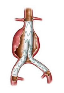
When elective surgery is recommended, three main approaches may be used:
- Open surgery, which involves an abdominal incision to replace the dilated section of the aorta with a synthetic graft.
- Endovascular repair, performed using a catheter inserted through an artery in the leg to place a stent graft inside the vessel without opening the abdomen.
- Hybrid technique, combining both methods depending on the aneurysm’s shape and location.
Consent Form
Before proceeding with the recommended intervention, an informed consent form must be signed.
This document confirms that the individual has received all the necessary information about the benefits, risks, and available alternatives related to the procedure.
In most cases, these aspects have already been discussed and all questions have been answered.
At this stage, the physician considers that the expected benefits of the procedure outweigh its potential risks.

-Open Surgery
Open surgery involves replacing the dilated portion of the aorta with a synthetic tube, most often made of Dacron.
It is a major procedure, usually recommended for younger patients in good physical condition.
The operation lasts about two to three hours, followed by a hospital stay of five to ten days.
Recovery takes time, with a gradual return to normal daily activities typically between six and twelve weeks after the procedure.
When performed electively, meaning not in an emergency context, the surgical risk is relatively low—usually below 5%.

-Endovascular Repair
Endovascular repair is a less invasive approach than open surgery.
It consists of inserting an endoprosthesis (or stent graft) into the aorta using a catheter introduced through both groin arteries, via small incisions.
Once in place, the stent graft is deployed to isolate the aneurysm from bloodBlood is composed of red blood cells, white blood cells, platelets, and plasma. Red blood cells are responsible for transporting oxygen and carbon dioxide. White blood cells make up our immune defense system. Platelets contribute to blood flow, redirecting circulation inside the graft.
This reduces pressure on the weakened wall and lowers the risk of rupture.
Approximately 80% of people with an abdominal aortic aneurysm are eligible for this type of treatment.
The procedure is generally shorter than open surgery, lasting less than one hour, and hospital discharge often occurs the same day or the following day.
Recovery is usually quick, with a gradual return to activity within two to six weeks.
Regular imaging follow-up is essential to check the position of the stent graft and ensure there are no long-term complications.
In a planned, non-emergency context, the procedural risk remains low, generally under 3%.
-Hybrid Technique
The hybrid technique combines elements of open surgery and endovascular repair.
It is used when the aneurysm’s shape or location prevents the exclusive use of one approach.
The surgeon may repair part of the aorta through open surgery while placing a stent graft in another section.
This approach offers a tailored solution, adapted to each patient’s anatomy and overall condition.
The duration of the procedure, recovery, and follow-up depend on the complexity of the repair and the portion of the aorta involved.
Postoperative Follow-Up
After surgical or endovascular repair of the aorta, regular medical follow-up remains essential.
Imaging—whether ultrasound, CT scan, or MRI—helps monitor the condition of the rest of the aorta, detect possible complications, and adjust treatment when necessary.
Over time, other segments of the aorta may also dilate, sometimes requiring another intervention.
Long-term surveillance is therefore crucial to ensure stability of the repair and prevent recurrence.
Once healing has begun, daily activities can be gradually resumed.
Physical restrictions are lifted over time, but recovery should progress gradually and without excess.
The intensity and type of exercise are adjusted according to overall health, the type of intervention performed, and specific medical recommendations for each case.

Prevention
Prevention plays a key role in reducing the risk of developing or worsening an aortic aneurysm.
It relies primarily on the management of cardiovascular risk factors.
- Smoking cessation is the most effective measure to slow aneurysm progression. Smoking directly damages the arterial wall and accelerates its weakening.
- Controlling bloodBlood is composed of red blood cells, white blood cells, platelets, and plasma. Red blood cells are responsible for transporting oxygen and carbon dioxide. White blood cells make up our immune defense system. Platelets contribute to blood pressure and cholesterolCholesterol is essential for the proper functioning of the human body, but it can also have harmful effects if present in excess. >> reduces the strain on the vessel wall and limits local inflammation.
- A balanced diet, rich in fruits, vegetables, and fiber and low in saturated fats, helps maintain healthy cholesterolCholesterol is essential for the proper functioning of the human body, but it can also have harmful effects if present in excess. >> levels.
- Regular moderate physical activity supports vascular health, helps maintain a healthy weight, and assists in controlling bloodBlood is composed of red blood cells, white blood cells, platelets, and plasma. Red blood cells are responsible for transporting oxygen and carbon dioxide. White blood cells make up our immune defense system. Platelets contribute to blood pressure and diabetes.
Regular medical monitoring complements these measures, allowing for treatment adjustments, surveillance of aortic size, and early detection of any progression.
Prevention therefore relies on a combination of lifestyle habits and medical follow-up that together help reduce the risk of serious complications associated with aneurysms.
Read more: Healthy lifestyle habits
Conclusion
When an abdominal aortic aneurysm is discovered—often incidentally—the main goal is to prevent rupture or tearing of the wall, events that can be life-threatening.
The true challenge lies in determining the right moment to intervene, when the benefits of surgery outweigh the risks of waiting.
Close monitoring and individualized management by a specialized vascular surgery team help anticipate this decision and act before complications occur.
Related articles


Latest articles