The heart has two valves located between the atriaThe atria are the two upper chambers of the heart. They act as reservoirs for blood that will fill the ventricles. and the ventricles. Their role is simple yet essential: they open to allow bloodBlood is composed of red blood cells, white blood cells, platelets, and plasma. Red blood cells are responsible for transporting oxygen and carbon dioxide. White blood cells make up our immune defense system. Platelets contribute to blood to pass through and close to prevent it from flowing backward, thereby ensuring efficient, one-way circulation.
On the right side is the tricuspid valve, and on the left side, the mitral valve.
The mitral valve is not simply a thin membrane that opens and closes. It is a true mechanical system known as the mitral apparatus. It includes a sturdy ring (the annulus), two mobile leaflets, chordae tendineae, and papillary muscles located deep within the left ventricle.
All these structures work in coordination to allow the valve to close perfectly with each heartbeat. This harmony ensures a tight seal between the left ventricle and the left atrium.
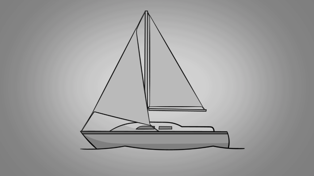
A Simple Comparison: The Sailboat
To better understand, the mitral apparatus can be compared to a sailboat. The fibrous annulus acts as the mast—the solid structure to which the sail is attached at the top. The sail corresponds to the two mitral valve leaflets.
From the edges of the leaflets, numerous chordae extend downward and attach to the papillary muscles at the base of the left ventricle. These muscles serve as anchor points that keep the sail properly tensioned.
In this way, the chordae prevent the leaflets from billowing or flipping backward under bloodBlood is composed of red blood cells, white blood cells, platelets, and plasma. Red blood cells are responsible for transporting oxygen and carbon dioxide. White blood cells make up our immune defense system. Platelets contribute to blood pressure, much like they prevent a sail from flapping in the wind and losing efficiency.
Thanks to this well-organized structure, the valve closes in a stable manner and minimizes leakage into the left atrium.
Ventricular Contraction
The heart’s electrical system triggers ventricular contraction from the bottom upward. Pressure rises rapidly within the chamber, forcing the two mitral valve leaflets to close.
Normal Closure of the Mitral Valve
When the mitral valve closes properly, the edges of the two leaflets come together and seal firmly against each other.
The chordae tendineae keep the leaflets taut and prevent them from folding back into the left atrium. In this way, the valve forms an effective barrier: bloodBlood is composed of red blood cells, white blood cells, platelets, and plasma. Red blood cells are responsible for transporting oxygen and carbon dioxide. White blood cells make up our immune defense system. Platelets contribute to blood can no longer flow backward.
The only possible pathway is then through the aortic valveThe aortic valve is located between the left ventricule and the aorta. It is one of the four valves ot the heart. >>, allowing bloodBlood is composed of red blood cells, white blood cells, platelets, and plasma. Red blood cells are responsible for transporting oxygen and carbon dioxide. White blood cells make up our immune defense system. Platelets contribute to blood to continue forward circulation.
Significant Pressure on the Mitral Valve
The tension applied to the mitral valve is naturally very high with each heartbeat. It becomes even greater in individuals with high bloodBlood is composed of red blood cells, white blood cells, platelets, and plasma. Red blood cells are responsible for transporting oxygen and carbon dioxide. White blood cells make up our immune defense system. Platelets contribute to blood pressure.
Imagine the force the mitral apparatus must withstand when pressure can exceed 200 millimeters of mercury (mm Hg)!
When This Mechanism Fails
For the mitral valve to remain perfectly sealed, each component of the mitral apparatus must function properly: the fibrous annulus, the two leaflets, the chordae tendineae, and the papillary muscles.
If any of these elements deteriorates, becomes deformed, or loses mobility, closure becomes imperfect. A small gap may then appear between the leaflets, allowing part of the bloodBlood is composed of red blood cells, white blood cells, platelets, and plasma. Red blood cells are responsible for transporting oxygen and carbon dioxide. White blood cells make up our immune defense system. Platelets contribute to blood to flow back into the left atrium. This condition is known as mitral regurgitation.
This leakage may occur acutely (suddenly) or develop gradually over time in a chronic (progressive) form.
The mitral valve can also be affected by another type of disorder: narrowing of its opening, known as mitral stenosis. Unlike mitral regurgitation, where the valve does not close properly, stenosis prevents bloodBlood is composed of red blood cells, white blood cells, platelets, and plasma. Red blood cells are responsible for transporting oxygen and carbon dioxide. White blood cells make up our immune defense system. Platelets contribute to blood from flowing freely from the left atrium into the left ventricle.
Today, the main cause of this structural change is acute rheumatic fever, a complication of an inadequately treated streptococcal infection. This condition can lead to thickening and stiffening of the leaflets, sometimes accompanied by partial fusion of the supporting structures.
Although a “pure” form of mitral stenosis can exist, it is often associated with leakage of varying severity. In such cases, it is referred to as mixed mitral valve disease, combining both obstruction to forward flow and incomplete closure.