Congenital aortic stenosis refers to a narrowing of the aortic valveThe aortic valve is located between the left ventricule and the aorta. It is one of the four valves ot the heart. >> that is present from birth. The degree of narrowing varies from one person to another and determines both the frequency of follow-up and the available treatment options.
What Is It?
The aortic valveThe aortic valve is located between the left ventricule and the aorta. It is one of the four valves ot the heart. >> acts as the heart’s exit door. It opens to allow bloodBlood is composed of red blood cells, white blood cells, platelets, and plasma. Red blood cells are responsible for transporting oxygen and carbon dioxide. White blood cells make up our immune defense system. Platelets contribute to blood to leave the left ventricle and enter the aorta, the large artery responsible for distributing oxygenated bloodBlood is composed of red blood cells, white blood cells, platelets, and plasma. Red blood cells are responsible for transporting oxygen and carbon dioxide. White blood cells make up our immune defense system. Platelets contribute to blood throughout the body. When this opening is too narrow, the heart must work harder to push bloodBlood is composed of red blood cells, white blood cells, platelets, and plasma. Red blood cells are responsible for transporting oxygen and carbon dioxide. White blood cells make up our immune defense system. Platelets contribute to blood forward.
Opening and Closing the Aortic Valve
The heart functions in a cycle made up of two phases: a filling phase, during which the heart muscle relaxes, and a contraction phase, during which it ejects bloodBlood is composed of red blood cells, white blood cells, platelets, and plasma. Red blood cells are responsible for transporting oxygen and carbon dioxide. White blood cells make up our immune defense system. Platelets contribute to blood.
When the left ventricle contracts and the pressure inside the heart exceeds that of the aorta, the aortic valveThe aortic valve is located between the left ventricule and the aorta. It is one of the four valves ot the heart. >> opens, allowing bloodBlood is composed of red blood cells, white blood cells, platelets, and plasma. Red blood cells are responsible for transporting oxygen and carbon dioxide. White blood cells make up our immune defense system. Platelets contribute to blood to be expelled into the artery.
Once this task is completed, the heart relaxes and enters its resting phase. The bloodBlood is composed of red blood cells, white blood cells, platelets, and plasma. Red blood cells are responsible for transporting oxygen and carbon dioxide. White blood cells make up our immune defense system. Platelets contribute to blood within the aorta then naturally tends to flow back toward the ventricle as pressure falls. This movement causes the leaflets of the aortic valveThe aortic valve is located between the left ventricule and the aorta. It is one of the four valves ot the heart. >> to close immediately, preventing bloodBlood is composed of red blood cells, white blood cells, platelets, and plasma. Red blood cells are responsible for transporting oxygen and carbon dioxide. White blood cells make up our immune defense system. Platelets contribute to blood from flowing back into the heart.
This mechanism can be visualized as inverted parachutes that deploy to block backward flow.
Narrowing of the Aortic Valve
When the valve opening is too narrow, the heart must work harder to maintain adequate bloodBlood is composed of red blood cells, white blood cells, platelets, and plasma. Red blood cells are responsible for transporting oxygen and carbon dioxide. White blood cells make up our immune defense system. Platelets contribute to blood circulation. Over time, this extra effort can lead to fatigue of the heart muscle.
The narrowing may be mild, moderate, or severe, depending on the structure of the valve and the resistance it creates to bloodBlood is composed of red blood cells, white blood cells, platelets, and plasma. Red blood cells are responsible for transporting oxygen and carbon dioxide. White blood cells make up our immune defense system. Platelets contribute to blood flow.
In congenital forms, the abnormality is present at birth and results from altered valve development during fetal life. Symptoms may appear very early, or conversely, remain silent for many years, depending on the severity of the narrowing.
The Most Common Abnormality
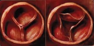
The most common congenital malformation of the aortic valveThe aortic valve is located between the left ventricule and the aorta. It is one of the four valves ot the heart. >> is the bicuspid aortic valveThe aortic valve is located between the left ventricule and the aorta. It is one of the four valves ot the heart. >>, meaning a valve made up of two leaflets instead of the usual three. It affects approximately 2% of the population.
In some individuals, the valve may even have only a single leaflet; this is referred to as a unicuspid valve. A bicuspid or unicuspid valve is not necessarily narrowed at birth, but it tends to thicken and calcify over time, which can gradually lead to aortic stenosis.
Other Possible Locations of the Narrowing
In some cases, the obstruction is not located at the valve itself, but rather just below or above it.
Below the valve (subaortic stenosis): Some individuals are born with a ridge or ring of tissue just beneath the aortic valveThe aortic valve is located between the left ventricule and the aorta. It is one of the four valves ot the heart. >>. This structure can interfere with bloodBlood is composed of red blood cells, white blood cells, platelets, and plasma. Red blood cells are responsible for transporting oxygen and carbon dioxide. White blood cells make up our immune defense system. Platelets contribute to blood flow when the heart contracts. The degree of obstruction may range from mild to severe, depending on the size of the abnormality.
Above the valve (supravalvular stenosis): This form is much rarer and is often associated with specific genetic syndromes.
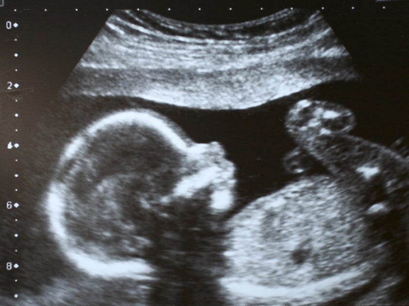
Prenatal Diagnosis
When aortic stenosis is severe, it can sometimes be detected before birth during routine fetal ultrasound examinations. Certain findings may raise suspicion, such as:
a dilated or thickened left ventricle,
limited movement of the aortic valveThe aortic valve is located between the left ventricule and the aorta. It is one of the four valves ot the heart. >> leaflets,
or abnormal bloodBlood is composed of red blood cells, white blood cells, platelets, and plasma. Red blood cells are responsible for transporting oxygen and carbon dioxide. White blood cells make up our immune defense system. Platelets contribute to blood flow across the valve.
In such situations, the physician may recommend a specialized fetal echocardiogram, performed by a team experienced in prenatal cardiology.
Why This Diagnosis Is Important
A diagnosis made before birth makes it possible to:
plan delivery in a center with access to a pediatric cardiology team;
prepare for early intervention if the stenosis is considered critical at birth;
inform and support parents by explaining the expected outlook and the steps of follow-up care.
Particular Features of Fetal Circulation
During fetal life, the lungs are not yet used for breathing. Instead, the placenta performs all vital exchanges: it supplies oxygen to the baby’s bloodBlood is composed of red blood cells, white blood cells, platelets, and plasma. Red blood cells are responsible for transporting oxygen and carbon dioxide. White blood cells make up our immune defense system. Platelets contribute to blood and removes carbon dioxide.
At this stage, only a small amount of bloodBlood is composed of red blood cells, white blood cells, platelets, and plasma. Red blood cells are responsible for transporting oxygen and carbon dioxide. White blood cells make up our immune defense system. Platelets contribute to blood is directed to the lungs to allow them to develop. Most of the bloodBlood is composed of red blood cells, white blood cells, platelets, and plasma. Red blood cells are responsible for transporting oxygen and carbon dioxide. White blood cells make up our immune defense system. Platelets contribute to blood flow bypasses the lungs through two natural pathways:
The foramen ovale, an opening between the two atriaThe atria are the two upper chambers of the heart. They act as reservoirs for blood that will fill the ventricles. of the heart, allows bloodBlood is composed of red blood cells, white blood cells, platelets, and plasma. Red blood cells are responsible for transporting oxygen and carbon dioxide. White blood cells make up our immune defense system. Platelets contribute to blood to pass directly from the right side to the left without going through the lungs.
The ductus arteriosus, a vessel connecting the pulmonary artery to the aorta, provides a second bypass route, allowing additional bloodBlood is composed of red blood cells, white blood cells, platelets, and plasma. Red blood cells are responsible for transporting oxygen and carbon dioxide. White blood cells make up our immune defense system. Platelets contribute to blood to reach the general circulation without passing through the lungs.
These mechanisms are essential before birth and normally close shortly afterward, once the newborn begins to breathe independently.
Birth: A Particularly Critical Period
In cases of severe aortic stenosis, birth represents a critical moment because the ductus arteriosus usually closes within the first hours of life.
Once this pathway closes, bloodBlood is composed of red blood cells, white blood cells, platelets, and plasma. Red blood cells are responsible for transporting oxygen and carbon dioxide. White blood cells make up our immune defense system. Platelets contribute to blood must travel exclusively through the normal route—namely through the aortic valveThe aortic valve is located between the left ventricule and the aorta. It is one of the four valves ot the heart. >> and the ascending aorta. If a significant narrowing is present, the left ventricle must generate a much greater effort to pump bloodBlood is composed of red blood cells, white blood cells, platelets, and plasma. Red blood cells are responsible for transporting oxygen and carbon dioxide. White blood cells make up our immune defense system. Platelets contribute to blood to the entire body. This can quickly lead to:
a deterioration in the newborn’s condition, such as pallor, rapid breathing, or low bloodBlood is composed of red blood cells, white blood cells, platelets, and plasma. Red blood cells are responsible for transporting oxygen and carbon dioxide. White blood cells make up our immune defense system. Platelets contribute to blood pressure;
reduced bloodBlood is composed of red blood cells, white blood cells, platelets, and plasma. Red blood cells are responsible for transporting oxygen and carbon dioxide. White blood cells make up our immune defense system. Platelets contribute to blood flow to the brain and the rest of the body;
a risk of acute heart failure.
For this reason, in severe cases identified before or immediately after birth, a medication called prostaglandin may be administered to temporarily keep the ductus arteriosus open. This helps stabilize the infant and allows time to plan the appropriate intervention.
Detection After Birth
When congenital aortic stenosis has not been identified before birth, it may become apparent later in life.
In newborns, a severe form often presents with a heart murmur and may be associated with breathing difficulties, poor skin coloration, or poor feeding.
During childhood or adolescence, the condition is frequently discovered incidentally during a routine medical examination, when the physician detects a heart murmur.
In adults, the diagnosis is sometimes made later, following the onset of symptoms during physical exertion, during the evaluation of a newly diagnosed arrhythmia, or during a medical examination performed for another reason.
Evolution
The course of congenital aortic stenosis varies depending on its type and severity. Mild narrowings—whether valvular, subvalvular, or supravalvular—may remain stable for many years, and sometimes for an entire lifetime.
When the narrowing is moderate or severe, the left ventricle gradually thickens to compensate for the increased workload, but this adaptive mechanism eventually reaches its limits. In some cases, the narrowing worsens over time or with growth, particularly if calcification develops in the valve or the aortic wall.
Without appropriate management, severe aortic stenosis may lead to heart failure, heart rhythm disturbances, or, more rarely, sudden death.
Follow Up
Congenital aortic stenosis requires lifelong medical follow-up.
The frequency of follow-up visits depends on the severity of the narrowing. Some patients undergo an echocardiogram every year, while others may be evaluated every two, three, or even five years, depending on the stability of their condition.
Precautions
-Physical Activity
In mild forms, most aerobic physical activities—such as walking, cycling, or swimming—are allowed. However, when the stenosis is moderate or severe, intense or competitive physical exertion is often discouraged.
Heavy weightlifting should be avoided regardless of severity, due to the potential fragility of the aortic wall. Sustained high-intensity effort may promote premature dilation of the aorta.
-Prevention of Valve Infection
Prevention of infective endocarditis relies primarily on good oral hygiene and regular dental care, including annual dental visits. Preventive antibiotics are no longer routinely required before dental procedures.
In women, any plan for pregnancy should be discussed in advance with a cardiologist. Severe, uncorrected aortic stenosis represents a contraindication to pregnancy because of the high risk of complications for both the mother and the baby.
-Family Screening
Family screening with echocardiography is recommended only in cases of a bicuspid aortic valveThe aortic valve is located between the left ventricule and the aorta. It is one of the four valves ot the heart. >>.
It applies to immediate family members—that is, parents, brothers, and sisters.
No routine screening is recommended for subvalvular or supravalvular forms of aortic stenosis.
Treatment
Treatment options depend on both the location of the stenosis and its degree of severity.
A – Valvular Forms
In mild to moderate valvular forms, careful medical follow-up is often sufficient for many years.
When the stenosis becomes importante, balloon valvuloplasty may be considered in children or adolescents. This procedure involves temporarily separating the valve leaflets using a small balloon inflated inside the valve. It can improve bloodBlood is composed of red blood cells, white blood cells, platelets, and plasma. Red blood cells are responsible for transporting oxygen and carbon dioxide. White blood cells make up our immune defense system. Platelets contribute to blood flow, but its effects are often temporary.
In adults, this technique is generally ineffective because the valve is usually too stiff and calcified.
1-Valve Replacement
In adults, valve replacement often becomes necessary. Several options are available:
Biological valve prosthesis, made from animal tissue
Mechanical valve prosthesis
Ross procedure, in which the patient’s own pulmonary valve is used to replace the diseased aortic valveThe aortic valve is located between the left ventricule and the aorta. It is one of the four valves ot the heart. >>, while the pulmonary valve is then replaced with a graft
The Ross procedure offers the advantage of providing a living valve that can grow with the child and preserves natural valve function without the need for lifelong anticoagulation. However, it creates two operated valve sites, which may require future reintervention, particularly to replace the pulmonary graft.
2-Other Approaches
Transcatheter aortic valveThe aortic valve is located between the left ventricule and the aorta. It is one of the four valves ot the heart. >> implantation (TAVI), widely used in older adults, is rarely indicated in congenital forms of aortic stenosis. The anatomy of congenital valves is often too complex or atypical for this type of procedure.
B – Subvalvular Forms
For subvalvular aortic stenosis, surgery primarily aims to remove the fibrous membrane or to reshape the left ventricular outflow tract in order to restore normal bloodBlood is composed of red blood cells, white blood cells, platelets, and plasma. Red blood cells are responsible for transporting oxygen and carbon dioxide. White blood cells make up our immune defense system. Platelets contribute to blood flow.
C – Supravalvular Forms
Supravalvular aortic stenosis, which is less common, may require surgical intervention to enlarge the initial portion of the aorta.
Balancing Risks and Benefits
Regardless of the type of stenosis, the decision to proceed with treatment is based on a careful balance between the expected benefits, the risks of the intervention, and the patient’s overall condition. Ongoing follow-up with a cardiologist is essential to determine the most appropriate timing for intervention.
In Summary
Congenital aortic stenosis may involve the valve itself, the region just below it, or just above it. When the obstruction is severe, it can sometimes be detected before birth, allowing for optimal management from the very first moments of life.
Regular and individualized follow-up is essential to preserve heart function and prevent complications.